6R1P

EthR ligand complex


Experimental Data Snapshot

  • Method: X-RAY DIFFRACTION
  • Resolution: 1.80 Å
  • R-Value Free: 0.205 
  • R-Value Work: 0.177 
  • R-Value Observed: 0.178 

wwPDB Validation   3D Report Full Report


Ligand Structure Quality Assessment 


This is version 1.1 of the entry. See complete history


Literature

Relative Binding Energies Predict Crystallographic Binding Modes of Ethionamide Booster Lead Compounds.

Tatum, N.J.Duarte, F.Kamerlin, S.C.L.Pohl, E.

(2019) J Phys Chem Lett 10: 2244-2249

  • DOI: https://doi.org/10.1021/acs.jpclett.9b00741
  • Primary Citation of Related Structures:  
    6R1P, 6R1S

  • PubMed Abstract: 

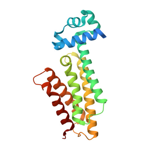
    Transcriptional repressor EthR from Mycobacterium tuberculosis is a valuable target for antibiotic booster drugs. We previously reported a virtual screening campaign to identify EthR inhibitors for development. Two ligand binding orientations were often proposed, though only the top scoring pose was utilized for filtering of the large data set. We obtained biophysically validated hits, some of which yielded complex crystal structures. In some cases, the crystallized binding mode and top scoring mode agree, while for others an alternate ligand binding orientation was found. In this contribution, we combine rigid docking, molecular dynamics simulations, and the linear interaction energy method to calculate binding free energies and derive relative binding energies for a number of EthR inhibitors in both modes. This strategy allowed us to correctly predict the most favorable orientation. Therefore, this widely applicable approach will be suitable to triage multiple binding modes within EthR and other potential drug targets with similar characteristics.


  • Organizational Affiliation

    Department of Chemistry , Durham University , South Road , Durham DH1 3LE , U.K.


Macromolecules
Find similar proteins by:  (by identity cutoff)  |  3D Structure
Entity ID: 1
MoleculeChains Sequence LengthOrganismDetailsImage
HTH-type transcriptional regulator EthR216Mycobacterium tuberculosisMutation(s): 0 
Gene Names: ethRetaRRv3855
UniProt
Find proteins for P9WMC1 (Mycobacterium tuberculosis (strain ATCC 25618 / H37Rv))
Explore P9WMC1 
Go to UniProtKB:  P9WMC1
Entity Groups  
Sequence Clusters30% Identity50% Identity70% Identity90% Identity95% Identity100% Identity
UniProt GroupP9WMC1
Sequence Annotations
Expand
  • Reference Sequence
Small Molecules
Ligands 1 Unique
IDChains Name / Formula / InChI Key2D Diagram3D Interactions
JPH
Query on JPH

Download Ideal Coordinates CCD File 
B [auth A]2-[2-[4-(2,3-dihydro-1,4-benzodioxin-6-yl)-1,2,3-triazol-1-yl]ethyl]-6-methyl-1~{H}-pyrimidin-4-one
C17 H17 N5 O3
HUPUXSMUAITIDA-UHFFFAOYSA-N
Experimental Data & Validation

Experimental Data

  • Method: X-RAY DIFFRACTION
  • Resolution: 1.80 Å
  • R-Value Free: 0.205 
  • R-Value Work: 0.177 
  • R-Value Observed: 0.178 
  • Space Group: P 41 21 2
Unit Cell:
Length ( Å )Angle ( ˚ )
a = 121.764α = 90
b = 121.764β = 90
c = 33.625γ = 90
Software Package:
Software NamePurpose
REFMACrefinement
XDSdata reduction
XSCALEdata scaling
PHASERphasing

Structure Validation

View Full Validation Report


Ligand Structure Quality Assessment 


Entry History 

Deposition Data

Revision History  (Full details and data files)

  • Version 1.0: 2020-02-19
    Type: Initial release
  • Version 1.1: 2024-01-24
    Changes: Data collection, Database references, Refinement description