


Structural basis for chemically-induced homodimerization of a single domain antibody
Lesne, J.x.To be published.
Experimental Data Snapshot
Starting Model: experimental
View more details
Entity ID: 1 | |||||
|---|---|---|---|---|---|
| Molecule | Chains | Sequence Length | Organism | Details | Image |
| VHH | A [auth H], B [auth A], E [auth D], F [auth E] | 119 | Camelidae | Mutation(s): 0 | 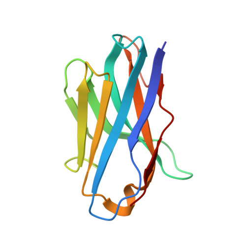 |
Entity ID: 2 | |||||
|---|---|---|---|---|---|
| Molecule | Chains | Sequence Length | Organism | Details | Image |
| VHH | C [auth B], D [auth C], G [auth F], H [auth G] | 120 | Camelidae | Mutation(s): 0 | 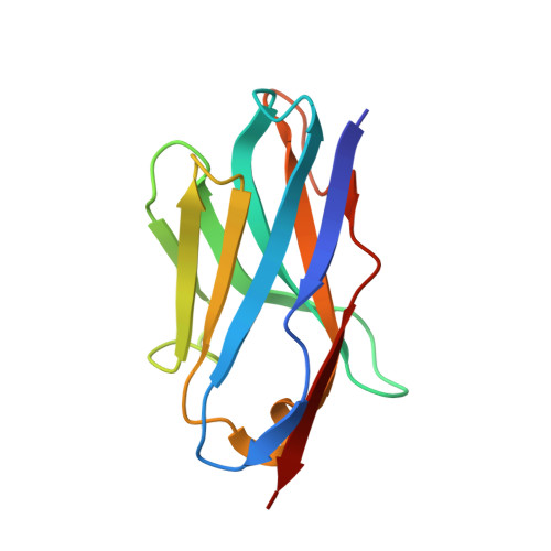 |
| Ligands 1 Unique | |||||
|---|---|---|---|---|---|
| ID | Chains | Name / Formula / InChI Key | 2D Diagram | 3D Interactions | |
| CFF Download:Ideal Coordinates CCD File | I [auth H], J [auth A], K [auth D], L [auth E] | CAFFEINE C8 H10 N4 O2 RYYVLZVUVIJVGH-UHFFFAOYSA-N |  | ||
| Length ( Å ) | Angle ( ˚ ) |
|---|---|
| a = 83.26 | α = 90 |
| b = 60.99 | β = 93.69 |
| c = 87.67 | γ = 90 |
| Software Name | Purpose |
|---|---|
| XDS | data reduction |
| XSCALE | data scaling |
| PHASER | phasing |
| PHENIX | refinement |
| PDB_EXTRACT | data extraction |


