Redox-induced structural changes in the di-iron and di-manganese forms of Bacillus anthracis ribonucleotide reductase subunit NrdF suggest a mechanism for gating of radical access.
Grave, K., Lambert, W., Berggren, G., Griese, J.J., Bennett, M.D., Logan, D.T., Hogbom, M.(2019) J Biol Inorg Chem 24: 849-861
- PubMed: 31410573 Search on PubMedSearch on PubMed Central
- DOI: https://doi.org/10.1007/s00775-019-01703-z
- Primary Citation Related Structures:
6QO5, 6QO7, 6QO8, 6QO9, 6QOB - PubMed Abstract:
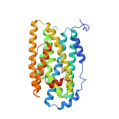
Class Ib ribonucleotide reductases (RNR) utilize a di-nuclear manganese or iron cofactor for reduction of superoxide or molecular oxygen, respectively. This generates a stable tyrosyl radical (Y·) in the R2 subunit (NrdF), which is further used for ribonucleotide reduction in the R1 subunit of RNR. Here, we report high-resolution crystal structures of Bacillus anthracis NrdF in the metal-free form (1.51 Å) and in complex with manganese (Mn II /Mn II , 1.30 Å). We also report three structures of the protein in complex with iron, either prepared anaerobically (Fe II /Fe II form, 1.32 Å), or prepared aerobically in the photo-reduced Fe II /Fe II form (1.63 Å) and with the partially oxidized metallo-cofactor (1.46 Å). The structures reveal significant conformational dynamics, likely to be associated with the generation, stabilization, and transfer of the radical to the R1 subunit. Based on observed redox-dependent structural changes, we propose that the passage for the superoxide, linking the FMN cofactor of NrdI and the metal site in NrdF, is closed upon metal oxidation, blocking access to the metal and radical sites. In addition, we describe the structural mechanics likely to be involved in this process.
- Department of Biochemistry and Biophysics, Stockholm University, Svante Arrhenius väg 16C, 10691, Stockholm, Sweden.
Organizational Affiliation: