


Three dimensional structure of human carbonic anhydrase XII in complex with benzenesulfonamide
Dvinskis, E., Leitans, J., Tars, K.To be published.
Experimental Data Snapshot
Starting Model: experimental
View more details
Entity ID: 1 | |||||
|---|---|---|---|---|---|
| Molecule | Chains | Sequence Length | Organism | Details | Image |
| Carbonic anhydrase 12 | 263 | Homo sapiens | Mutation(s): 0 Gene Names: CA12 EC: 4.2.1.1 | 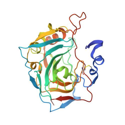 | |
UniProt & NIH Common Fund Data Resources | |||||
PHAROS: O43570 GTEx: ENSG00000074410 | |||||
Entity Groups | |||||
| Sequence Clusters | 30% Identity50% Identity70% Identity90% Identity95% Identity100% Identity | ||||
| UniProt Group | O43570 | ||||
Sequence AnnotationsExpand | |||||
Reference Sequence | |||||
| Ligands 2 Unique | |||||
|---|---|---|---|---|---|
| ID | Chains | Name / Formula / InChI Key | 2D Diagram | 3D Interactions | |
| J95 (Subject of Investigation/LOI) Download:Ideal Coordinates CCD File | F [auth A], H [auth B], J [auth C], L [auth D] | ~{N}-butyl-4-chloranyl-2-(cyclohexylamino)-5-sulfamoyl-benzamide C17 H26 Cl N3 O3 S WVNMCWQJUJCKNS-UHFFFAOYSA-N |  | ||
| ZN Download:Ideal Coordinates CCD File | E [auth A], G [auth B], I [auth C], K [auth D] | ZINC ION Zn PTFCDOFLOPIGGS-UHFFFAOYSA-N |  | ||
| Length ( Å ) | Angle ( ˚ ) |
|---|---|
| a = 46.93 | α = 82.09 |
| b = 66.05 | β = 84.41 |
| c = 80.95 | γ = 86.17 |
| Software Name | Purpose |
|---|---|
| REFMAC | refinement |
| SCALA | data scaling |


