6QML | pdb_00006qml

UCHL3 in complex with synthetic, K27-linked diubiquitin


Experimental Data Snapshot

  • Method: X-RAY DIFFRACTION
  • Resolution: 2.10 Å
  • R-Value Free: 
    0.233 (Depositor), 0.240 (DCC) 
  • R-Value Work: 
    0.184 (Depositor), 0.195 (DCC) 
  • R-Value Observed: 
    0.187 (Depositor) 

Starting Models: experimental
View more details

wwPDB Validation 3D Report Full Report

Validation slider image for 6QML

This is version 1.4 of the entry. See complete history

Literature

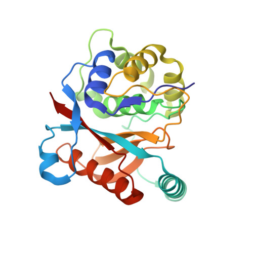
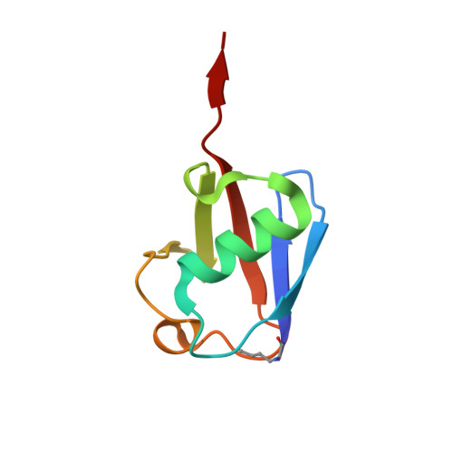
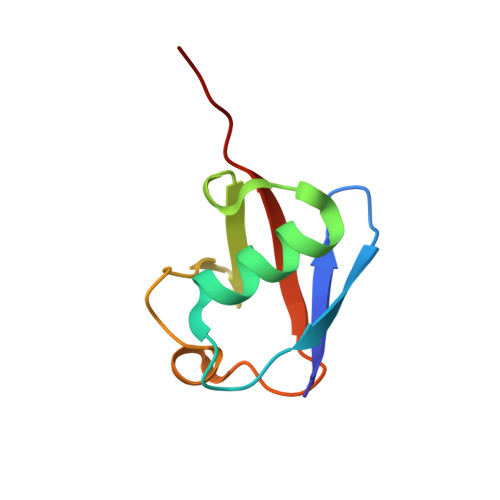
K27-Linked Diubiquitin Inhibits UCHL3 via an Unusual Kinetic Trap.

van Tilburg, G.B.A.Murachelli, A.G.Fish, A.van der Heden van Noort, G.J.Ovaa, H.Sixma, T.K.

(2021) Cell Chem Biol 28: 191-201.e8

  • DOI: https://doi.org/10.1016/j.chembiol.2020.11.005
  • Primary Citation Related Structures: 
    6QML

  • PubMed Abstract: 

    Functional analysis of lysine 27-linked ubiquitin chains ( K27 Ub) is difficult due to the inability to make them through enzymatic methods and due to a lack of model tools and substrates. Here we generate a series of ubiquitin (Ub) tools to study how the deubiquitinase UCHL3 responds to K27 Ub chains in comparison to lysine 63-linked chains and mono-Ub. From a crystal structure of a complex between UCHL3 and synthetic K27 Ub 2 , we unexpectedly discover that free K27 Ub 2 and K27 Ub 2 -conjugated substrates are natural inhibitors of UCHL3. Using our Ub tools to profile UCHL3's activity, we generate a quantitative kinetic model of the inhibitory mechanism and we find that K27 Ub 2 can inhibit UCHL3 covalently, by binding to its catalytic cysteine, and allosterically, by locking its catalytic loop tightly in place. Based on this inhibition mechanism, we propose that UCHL3 and K27 Ub chains likely sense and regulate each other in cells.


  • Organizational Affiliation
    • Department of Cell and Chemical Biology and Oncode Institute, Leiden University Medical Center, Einthovenweg 20, 2333 ZC Leiden, the Netherlands.

Macromolecule Content 

  • Total Structure Weight: 87.58 kDa 
  • Atom Count: 6,397 
  • Modeled Residue Count: 758 
  • Deposited Residue Count: 758 
  • Unique protein chains: 3

Macromolecules

Find similar proteins by:|  3D Structure
Entity ID: 1
MoleculeChains  Sequence LengthOrganismDetailsImage
Ubiquitin carboxyl-terminal hydrolase isozyme L3
A, D
227Homo sapiensMutation(s): 0 
Gene Names: UCHL3
EC: 3.4.19.12
UniProt & NIH Common Fund Data Resources
Find proteins for P15374 (Homo sapiens)
Explore P15374 
Go to UniProtKB:  P15374
PHAROS:  P15374
GTEx:  ENSG00000118939 
Entity Groups
Sequence Clusters30% Identity50% Identity70% Identity90% Identity95% Identity100% Identity
UniProt GroupP15374
Sequence Annotations
Expand
Reference Sequence
Find similar proteins by:|  3D Structure
Entity ID: 2
MoleculeChains  Sequence LengthOrganismDetailsImage
Polyubiquitin-B
B, E
76Homo sapiensMutation(s): 1 
UniProt & NIH Common Fund Data Resources
Find proteins for P0CG47 (Homo sapiens)
Explore P0CG47 
Go to UniProtKB:  P0CG47
PHAROS:  P0CG47
GTEx:  ENSG00000170315 
Entity Groups
Sequence Clusters30% Identity50% Identity70% Identity90% Identity95% Identity100% Identity
UniProt GroupP0CG47
Sequence Annotations
Expand
Reference Sequence
Find similar proteins by:|  3D Structure
Entity ID: 3
MoleculeChains  Sequence LengthOrganismDetailsImage
Polyubiquitin-C
C, F
76Homo sapiensMutation(s): 0 
UniProt & NIH Common Fund Data Resources
Find proteins for P0CG48 (Homo sapiens)
Explore P0CG48 
Go to UniProtKB:  P0CG48
PHAROS:  P0CG48
GTEx:  ENSG00000150991 
Entity Groups
Sequence Clusters30% Identity50% Identity70% Identity90% Identity95% Identity100% Identity
UniProt GroupP0CG48
Sequence Annotations
Expand
Reference Sequence

Small Molecules

Ligands 4 Unique
IDChains Name / Formula / InChI Key2D Diagram3D Interactions
BR

Query on BR


Download:Ideal Coordinates CCD File
DA [auth F],
I [auth A],
O [auth B],
R [auth C]
BROMIDE ION
Br
CPELXLSAUQHCOX-UHFFFAOYSA-M
EDO

Query on EDO


Download:Ideal Coordinates CCD File
AA [auth D]
BA [auth D]
EA [auth F]
FA [auth F]
J [auth A]
AA [auth D],
BA [auth D],
EA [auth F],
FA [auth F],
J [auth A],
K [auth A],
L [auth A],
M [auth A],
P [auth B],
Q [auth B],
S [auth C],
T [auth C],
U [auth C],
Y [auth D],
Z [auth D]
1,2-ETHANEDIOL
C2 H6 O2
LYCAIKOWRPUZTN-UHFFFAOYSA-N
K

Query on K


Download:Ideal Coordinates CCD File
G [auth A],
H [auth A],
V [auth D],
W [auth D],
X [auth D]
POTASSIUM ION
K
NPYPAHLBTDXSSS-UHFFFAOYSA-N
CL

Query on CL


Download:Ideal Coordinates CCD File
CA [auth D],
N [auth A]
CHLORIDE ION
Cl
VEXZGXHMUGYJMC-UHFFFAOYSA-M
Modified Residues  1 Unique
IDChains TypeFormula2D DiagramParent
NLE
Query on NLE
B, E
L-PEPTIDE LINKINGC6 H13 N O2LEU

Experimental Data & Validation

Experimental Data

  • Method: X-RAY DIFFRACTION
  • Resolution: 2.10 Å
  • R-Value Free:  0.233 (Depositor), 0.240 (DCC) 
  • R-Value Work:  0.184 (Depositor), 0.195 (DCC) 
  • R-Value Observed: 0.187 (Depositor) 
Space Group: P 2 21 21
Unit Cell:
Length ( Å )Angle ( ˚ )
a = 70.29α = 90
b = 81.206β = 90
c = 159.858γ = 90
Software Package:
Software NamePurpose
REFMACrefinement
XDSdata reduction
SCALAdata scaling
PHASERphasing

Structure Validation

View Full Validation Report


Entry History 

& Funding Information

Deposition Data


Funding OrganizationLocationGrant Number
Oncode InstituteNetherlands--
CancerGenomiCs.nlNetherlands--

Revision History  (Full details and data files)

  • Version 1.0: 2020-02-26
    Type: Initial release
  • Version 1.1: 2020-05-20
    Changes: Advisory, Derived calculations
  • Version 1.2: 2021-03-10
    Changes: Database references, Derived calculations
  • Version 1.3: 2024-01-24
    Changes: Data collection, Database references, Refinement description
  • Version 1.4: 2024-10-23
    Changes: Structure summary