


Successful development of 3-oxohexahydrofuropyrrole amino acid amides as inhibitors of Cathepsin-K.
Derbyshire, D.J.To be published.
Experimental Data Snapshot
Entity ID: 1 | |||||
|---|---|---|---|---|---|
| Molecule | Chains | Sequence Length | Organism | Details | Image |
| Cathepsin K | 217 | Homo sapiens | Mutation(s): 0 Gene Names: CTSK, CTSO, CTSO2 EC: 3.4.22.38 | 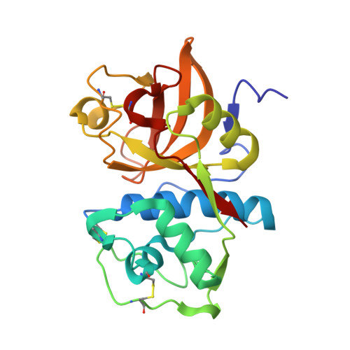 | |
UniProt & NIH Common Fund Data Resources | |||||
PHAROS: P43235 GTEx: ENSG00000143387 | |||||
Entity Groups | |||||
| Sequence Clusters | 30% Identity50% Identity70% Identity90% Identity95% Identity100% Identity | ||||
| UniProt Group | P43235 | ||||
Sequence AnnotationsExpand | |||||
Reference Sequence | |||||
| Ligands 2 Unique | |||||
|---|---|---|---|---|---|
| ID | Chains | Name / Formula / InChI Key | 2D Diagram | 3D Interactions | |
| J5N (Subject of Investigation/LOI) Download:Ideal Coordinates CCD File | B [auth A] | ~{N}-[(2~{S})-1-[(3~{R},3~{a}~{R},6~{R},6~{a}~{R})-6-ethynyl-3-oxidanyl-2,3,3~{a},5,6,6~{a}-hexahydrofuro[3,2-b]pyrrol-4-yl]-4-methyl-1-oxidanylidene-pentan-2-yl]-4-[5-fluoranyl-2-(4-methylpiperazin-1-yl)-1,3-thiazol-4-yl]benzamide C29 H36 F N5 O4 S LXQZJMLAONLFAB-KFGZMKLQSA-N |  | ||
| NO3 Download:Ideal Coordinates CCD File | C [auth A] D [auth A] E [auth A] F [auth A] G [auth A] | NITRATE ION N O3 NHNBFGGVMKEFGY-UHFFFAOYSA-N |  | ||
| Length ( Å ) | Angle ( ˚ ) |
|---|---|
| a = 70.91 | α = 90 |
| b = 70.91 | β = 90 |
| c = 54.27 | γ = 90 |
| Software Name | Purpose |
|---|---|
| MOSFLM | data reduction |
| Aimless | data scaling |
| PHASER | phasing |
| REFMAC | refinement |
| PDB_EXTRACT | data extraction |


