6QI1 | pdb_00006qi1

Time resolved structural analysis of the full turnover of an enzyme - 12312 ms


Experimental Data Snapshot

  • Method: X-RAY DIFFRACTION
  • Resolution: 1.90 Å
  • R-Value Free: 
    0.259 (Depositor), 0.259 (DCC) 
  • R-Value Work: 
    0.212 (Depositor), 0.212 (DCC) 
  • R-Value Observed: 
    0.214 (Depositor) 

wwPDB Validation 3D Report Full Report

Validation slider image for 6QI1

This is version 1.1 of the entry. See complete history

Literature

Time-resolved crystallography reveals allosteric communication aligned with molecular breathing.

Mehrabi, P.Schulz, E.C.Dsouza, R.Muller-Werkmeister, H.M.Tellkamp, F.Miller, R.J.D.Pai, E.F.

(2019) Science 365: 1167-1170

  • DOI: https://doi.org/10.1126/science.aaw9904
  • Primary Citation Related Structures: 
    6QHP, 6QHQ, 6QHS, 6QHT, 6QHU, 6QHV, 6QHW, 6QHX, 6QHY, 6QHZ, 6QI0, 6QI1, 6QI2, 6QI3

  • PubMed Abstract: 

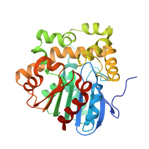
    A comprehensive understanding of protein function demands correlating structure and dynamic changes. Using time-resolved serial synchrotron crystallography, we visualized half-of-the-sites reactivity and correlated molecular-breathing motions in the enzyme fluoroacetate dehalogenase. Eighteen time points from 30 milliseconds to 30 seconds cover four turnover cycles of the irreversible reaction. They reveal sequential substrate binding, covalent-intermediate formation, setup of a hydrolytic water molecule, and product release. Small structural changes of the protein mold and variations in the number and placement of water molecules accompany the various chemical steps of catalysis. Triggered by enzyme-ligand interactions, these repetitive changes in the protein framework's dynamics and entropy constitute crucial components of the catalytic machinery.


  • Organizational Affiliation
    • Department for Atomically Resolved Dynamics, Max-Planck-Institute for Structure and Dynamics of Matter, Luruper Chaussee 149, 22761 Hamburg, Germany.

Macromolecule Content 

  • Total Structure Weight: 68.23 kDa 
  • Atom Count: 5,179 
  • Modeled Residue Count: 591 
  • Deposited Residue Count: 612 
  • Unique protein chains: 1

Macromolecules

Find similar proteins by:|  3D Structure
Entity ID: 1
MoleculeChains  Sequence LengthOrganismDetailsImage
Fluoroacetate dehalogenase
A, B
306Rhodopseudomonas palustrisMutation(s): 0 
Gene Names: RPA1163
EC: 3.8.1.3
UniProt
Find proteins for Q6NAM1 (Rhodopseudomonas palustris (strain ATCC BAA-98 / CGA009))
Explore Q6NAM1 
Go to UniProtKB:  Q6NAM1
Entity Groups
Sequence Clusters30% Identity50% Identity70% Identity90% Identity95% Identity100% Identity
UniProt GroupQ6NAM1
Sequence Annotations
Expand
Reference Sequence

Small Molecules

Ligands 1 Unique
IDChains Name / Formula / InChI Key2D Diagram3D Interactions
FAH

Query on FAH


Download:Ideal Coordinates CCD File
C [auth A]fluoroacetic acid
C2 H3 F O2
QEWYKACRFQMRMB-UHFFFAOYSA-N

Experimental Data & Validation

Experimental Data

  • Method: X-RAY DIFFRACTION
  • Resolution: 1.90 Å
  • R-Value Free:  0.259 (Depositor), 0.259 (DCC) 
  • R-Value Work:  0.212 (Depositor), 0.212 (DCC) 
  • R-Value Observed: 0.214 (Depositor) 
Space Group: P 1 21 1
Unit Cell:
Length ( Å )Angle ( ˚ )
a = 41.76α = 90
b = 79.61β = 102.86
c = 84.43γ = 90
Software Package:
Software NamePurpose
PHENIXrefinement
XDSdata reduction
XSCALEdata scaling
PHASERphasing

Structure Validation

View Full Validation Report


Entry History 

Deposition Data

Revision History  (Full details and data files)

  • Version 1.0: 2019-09-25
    Type: Initial release
  • Version 1.1: 2024-05-15
    Changes: Advisory, Data collection, Database references