A marine viral halogenase that iodinates diverse substrates.
Gkotsi, D.S., Ludewig, H., Sharma, S.V., Connolly, J.A., Dhaliwal, J., Wang, Y., Unsworth, W.P., Taylor, R.J.K., McLachlan, M.M.W., Shanahan, S., Naismith, J.H., Goss, R.J.M.(2019) Nat Chem 11: 1091-1097
- PubMed: 31611633 Search on PubMedSearch on PubMed Central
- DOI: https://doi.org/10.1038/s41557-019-0349-z
- Primary Citation Related Structures:
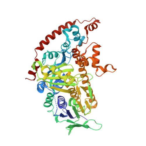
6QGM - PubMed Abstract:
Oceanic cyanobacteria are the most abundant oxygen-generating phototrophs on our planet and are therefore important to life. These organisms are infected by viruses called cyanophages, which have recently shown to encode metabolic genes that modulate host photosynthesis, phosphorus cycling and nucleotide metabolism. Herein we report the characterization of a wild-type flavin-dependent viral halogenase (VirX1) from a cyanophage. Notably, halogenases have been previously associated with secondary metabolism, tailoring natural products. Exploration of this viral halogenase reveals it capable of regioselective halogenation of a diverse range of substrates with a preference for forming aryl iodide species; this has potential implications for the metabolism of the infected host. Until recently, a flavin-dependent halogenase that is capable of iodination in vitro had not been reported. VirX1 is interesting from a biocatalytic perspective as it shows strikingly broad substrate flexibility and a clear preference for iodination, as illustrated by kinetic analysis. These factors together render it an attractive tool for synthesis.
- School of Chemistry, University of St Andrews, North Haugh, St Andrews, Fife, UK.
Organizational Affiliation: