6QEL | pdb_00006qel

E. coli DnaBC apo complex


Experimental Data Snapshot

  • Method: ELECTRON MICROSCOPY
  • Resolution: 3.90 Å
  • Aggregation State: PARTICLE 
  • Reconstruction Method: SINGLE PARTICLE 

wwPDB Validation 3D Report Full Report

Validation slider image for 6QEL

This is version 1.5 of the entry. See complete history

Literature

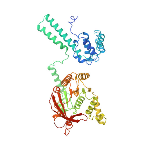
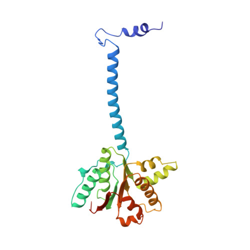
Physical Basis for the Loading of a Bacterial Replicative Helicase onto DNA.

Arias-Palomo, E.Puri, N.O'Shea Murray, V.L.Yan, Q.Berger, J.M.

(2019) Mol Cell 74: 173-184.e4

  • DOI: https://doi.org/10.1016/j.molcel.2019.01.023
  • Primary Citation Related Structures: 
    6QEL, 6QEM

  • PubMed Abstract: 

    In cells, dedicated AAA+ ATPases deposit hexameric, ring-shaped helicases onto DNA to initiate chromosomal replication. To better understand the mechanisms by which helicase loading can occur, we used cryo-EM to determine sub-4-Å-resolution structures of the E. coli DnaB⋅DnaC helicase⋅loader complex with nucleotide in pre- and post-DNA engagement states. In the absence of DNA, six DnaC protomers latch onto and crack open a DnaB hexamer using an extended N-terminal domain, stabilizing this conformation through nucleotide-dependent ATPase interactions. Upon binding DNA, DnaC hydrolyzes ATP, allowing DnaB to isomerize into a topologically closed, pre-translocation state competent to bind primase. Our data show how DnaC opens the DnaB ring and represses the helicase prior to DNA binding and how DnaC ATPase activity is reciprocally regulated by DnaB and DNA. Comparative analyses reveal how the helicase loading mechanism of DnaC parallels and diverges from homologous AAA+ systems involved in DNA replication and transposition.


  • Organizational Affiliation
    • Department of Structural & Chemical Biology, Centro de Investigaciones Biológicas, CIB-CSIC 28040 Madrid, Spain. Electronic address: earias@cib.csic.es.

Macromolecule Content 

  • Total Structure Weight: 488.29 kDa 
  • Atom Count: 32,514 
  • Modeled Residue Count: 4,092 
  • Deposited Residue Count: 4,296 
  • Unique protein chains: 2

Macromolecules

Find similar proteins by:|  3D Structure
Entity ID: 1
MoleculeChains  Sequence LengthOrganismDetailsImage
Replicative DNA helicase
A, B, C, D, E
A, B, C, D, E, F
471Escherichia coliMutation(s): 0 
Gene Names: A1UM_04839
EC: 3.6.4.12 (PDB Primary Data), 5.6.2.3 (UniProt)
UniProt
Find proteins for P0ACB0 (Escherichia coli (strain K12))
Explore P0ACB0 
Go to UniProtKB:  P0ACB0
Entity Groups
Sequence Clusters30% Identity50% Identity70% Identity90% Identity95% Identity100% Identity
UniProt GroupP0ACB0
Sequence Annotations
Expand
Reference Sequence
Find similar proteins by:|  3D Structure
Entity ID: 2
MoleculeChains  Sequence LengthOrganismDetailsImage
DNA replication protein dnaC
G, H, I, J, K
G, H, I, J, K, L
245Escherichia coliMutation(s): 0 
Gene Names: A1UM_00191
EC: 3.6.4
UniProt
Find proteins for P0AEF0 (Escherichia coli (strain K12))
Explore P0AEF0 
Go to UniProtKB:  P0AEF0
Entity Groups
Sequence Clusters30% Identity50% Identity70% Identity90% Identity95% Identity100% Identity
UniProt GroupP0AEF0
Sequence Annotations
Expand
Reference Sequence

Small Molecules

Ligands 3 Unique
IDChains Name / Formula / InChI Key2D Diagram3D Interactions
08T

Query on 08T


Download:Ideal Coordinates CCD File
AA [auth H],
CA [auth I],
EA [auth J],
GA [auth K],
IA [auth L]
[[[(2R,3S,4R,5R)-5-(6-aminopurin-9-yl)-3,4-bis(oxidanyl)oxolan-2-yl]methoxy-oxidanyl-phosphoryl]oxy-oxidanyl-phosphoryl]oxy-tris(fluoranyl)beryllium
C10 H14 Be F3 N5 O10 P2
WOGYHYSOODLXII-KWIZKVQNSA-J
ADP

Query on ADP


Download:Ideal Coordinates CCD File
M [auth A]
O [auth B]
Q [auth C]
S [auth D]
U [auth E]
M [auth A],
O [auth B],
Q [auth C],
S [auth D],
U [auth E],
W [auth F],
Y [auth G]
ADENOSINE-5'-DIPHOSPHATE
C10 H15 N5 O10 P2
XTWYTFMLZFPYCI-KQYNXXCUSA-N
MG

Query on MG


Download:Ideal Coordinates CCD File
BA [auth H]
DA [auth I]
FA [auth J]
HA [auth K]
JA [auth L]
BA [auth H],
DA [auth I],
FA [auth J],
HA [auth K],
JA [auth L],
N [auth A],
P [auth B],
R [auth C],
T [auth D],
V [auth E],
X [auth F],
Z [auth G]
MAGNESIUM ION
Mg
JLVVSXFLKOJNIY-UHFFFAOYSA-N

Experimental Data & Validation

Experimental Data

  • Method: ELECTRON MICROSCOPY
  • Resolution: 3.90 Å
  • Aggregation State: PARTICLE 
  • Reconstruction Method: SINGLE PARTICLE 
EM Software:
TaskSoftware PackageVersion
MODEL REFINEMENTPHENIX1.13
RECONSTRUCTIONRELION2.1

Structure Validation

View Full Validation Report


Entry History 

& Funding Information

Deposition Data


Funding OrganizationLocationGrant Number
United StatesR37-071747
SpainBFU2017-89143-P
SpainRYC-2015-19059

Revision History  (Full details and data files)

  • Version 1.0: 2019-03-06
    Type: Initial release
  • Version 1.1: 2019-04-17
    Changes: Data collection, Database references
  • Version 1.2: 2019-11-06
    Changes: Data collection, Refinement description
  • Version 1.3: 2019-12-04
    Changes: Derived calculations
  • Version 1.4: 2019-12-18
    Changes: Other
  • Version 1.5: 2024-05-15
    Changes: Data collection, Database references, Derived calculations