6QA7

Glycogen Phosphorylase b in complex with 29


Experimental Data Snapshot

  • Method: X-RAY DIFFRACTION
  • Resolution: 2.36 Å
  • R-Value Free: 0.191 
  • R-Value Work: 0.141 
  • R-Value Observed: 0.144 

wwPDB Validation   3D Report Full Report


Ligand Structure Quality Assessment 


This is version 1.2 of the entry. See complete history


Literature

Glucopyranosylidene-spiro-imidazolinones, a New Ring System: Synthesis and Evaluation as Glycogen Phosphorylase Inhibitors by Enzyme Kinetics and X-ray Crystallography.

Szabo, K.E.Kyriakis, E.Psarra, A.G.Karra, A.G.Sipos, A.Docsa, T.Stravodimos, G.A.Katsidou, E.Skamnaki, V.T.Liggri, P.G.V.Zographos, S.E.Mandi, A.Kiraly, S.B.Kurtan, T.Leonidas, D.D.Somsak, L.

(2019) J Med Chem 62: 6116-6136

  • DOI: https://doi.org/10.1021/acs.jmedchem.9b00356
  • Primary Citation of Related Structures:  
    6QA6, 6QA7, 6QA8

  • PubMed Abstract: 

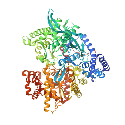
    Epimeric series of aryl-substituted glucopyranosylidene-spiro-imidazolinones, an unprecedented new ring system, were synthesized from the corresponding Schiff bases of O -perbenzoylated (gluculopyranosylamine)onamides by intramolecular ring closure of the aldimine moieties with the carboxamide group elicited by N -bromosuccinimide in pyridine. Test compounds were obtained by Zemplén O -debenzoylation. Stereochemistry and ring tautomers of the new compounds were investigated by NMR, time-dependent density functional theory (TDDFT)-electronic circular dichroism, and DFT-NMR methods. Kinetic studies with rabbit muscle and human liver glycogen phosphorylases showed that the ( R )-imidazolinones were 14-216 times more potent than the ( S ) epimers. The 2-naphthyl-substituted ( R )-imidazolinone was the best inhibitor of the human enzyme ( K i 1.7 μM) and also acted on HepG2 cells (IC 50 177 μM). X-ray crystallography revealed that only the ( R ) epimers bound in the crystal. Their inhibitory efficacy is based on the hydrogen-bonding interactions of the carbonyl oxygen and the NH of the imidazolinone ring.


  • Organizational Affiliation

    Department of Organic Chemistry , University of Debrecen , P.O. Box 400, H-4002 Debrecen , Hungary.


Macromolecules
Find similar proteins by:  (by identity cutoff)  |  3D Structure
Entity ID: 1
MoleculeChains Sequence LengthOrganismDetailsImage
Glycogen phosphorylase, muscle form843Oryctolagus cuniculusMutation(s): 0 
EC: 2.4.1.1
UniProt
Find proteins for P00489 (Oryctolagus cuniculus)
Explore P00489 
Go to UniProtKB:  P00489
Entity Groups  
Sequence Clusters30% Identity50% Identity70% Identity90% Identity95% Identity100% Identity
UniProt GroupP00489
Sequence Annotations
Expand
  • Reference Sequence
Small Molecules
Ligands 2 Unique
IDChains Name / Formula / InChI Key2D Diagram3D Interactions
HTW
Query on HTW

Download Ideal Coordinates CCD File 
B [auth A](5~{S},7~{R},8~{S},9~{S},10~{R})-7-(hydroxymethyl)-2-naphthalen-1-yl-8,9,10-tris(oxidanyl)-6-oxa-1,3-diazaspiro[4.5]dec-1-en-4-one
C18 H18 N2 O6
VIYRJCLPOZQRSZ-NSIVTLKISA-N
PLP
Query on PLP

Download Ideal Coordinates CCD File 
C [auth A]PYRIDOXAL-5'-PHOSPHATE
C8 H10 N O6 P
NGVDGCNFYWLIFO-UHFFFAOYSA-N
Binding Affinity Annotations 
IDSourceBinding Affinity
HTW Binding MOAD:  6QA7 Ki: 1.30e+4 (nM) from 1 assay(s)
Experimental Data & Validation

Experimental Data

  • Method: X-RAY DIFFRACTION
  • Resolution: 2.36 Å
  • R-Value Free: 0.191 
  • R-Value Work: 0.141 
  • R-Value Observed: 0.144 
  • Space Group: P 43 21 2
Unit Cell:
Length ( Å )Angle ( ˚ )
a = 128.212α = 90
b = 128.212β = 90
c = 115.944γ = 90
Software Package:
Software NamePurpose
REFMACrefinement
CrysalisProdata reduction
Aimlessdata scaling

Structure Validation

View Full Validation Report


Ligand Structure Quality Assessment 


Entry History 

Deposition Data

Revision History  (Full details and data files)

  • Version 1.0: 2019-06-26
    Type: Initial release
  • Version 1.1: 2019-07-10
    Changes: Data collection, Database references
  • Version 1.2: 2019-07-24
    Changes: Data collection, Database references