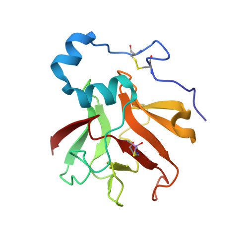
Structure and Electron-Transfer Pathway of the Human Methionine Sulfoxide Reductase MsrB3.
Javitt, G., Cao, Z., Resnick, E., Gabizon, R., Bulleid, N.J., Fass, D.(2020) Antioxid Redox Signal 33: 665-678
- PubMed: 32517586
- DOI: https://doi.org/10.1089/ars.2020.8037
- Primary Citation of Related Structures:
6Q9V, 6QA0 - PubMed Abstract:
Aims: The post-translational oxidation of methionine to methionine sulfoxide (MetSO) is a reversible process, enabling the repair of oxidative damage to proteins and the use of sulfoxidation as a regulatory switch. MetSO reductases catalyze the stereospecific reduction of MetSO. One of the mammalian MetSO reductases, MsrB3, has a signal sequence for entry into the endoplasmic reticulum (ER). In the ER, MsrB3 is expected to encounter a distinct redox environment compared with its paralogs in the cytosol, nucleus, and mitochondria. We sought to determine the location and arrangement of MsrB3 redox-active cysteines, which may couple MsrB3 activity to other redox events in the ER. Results: We determined the human MsrB3 structure by using X-ray crystallography. The structure revealed that a disulfide bond near the protein amino terminus is distant in space from the active site. Nevertheless, biochemical assays showed that these amino-terminal cysteines are oxidized by the MsrB3 active site after its reaction with MetSO. Innovation: This study reveals a mechanism to shuttle oxidizing equivalents from the primary MsrB3 active site toward the enzyme surface, where they would be available for further dithiol-disulfide exchange reactions. Conclusion: Conformational changes must occur during the MsrB3 catalytic cycle to transfer oxidizing equivalents from the active site to the amino-terminal redox-active disulfide. The accessibility of this exposed disulfide may help couple MsrB3 activity to other dithiol-disulfide redox events in the secretory pathway.
- Department of Structural Biology and Weizmann Institute of Science, Rehovot, Israel.
Organizational Affiliation:


