The structure of N184K amyloidogenic variant of gelsolin highlights the role of the H-bond network for protein stability and aggregation properties.
de Rosa, M., Barbiroli, A., Boni, F., Scalone, E., Mattioni, D., Vanoni, M.A., Patrone, M., Bollati, M., Mastrangelo, E., Giorgino, T., Milani, M.(2020) Eur Biophys J 49: 11-19
- PubMed: 31724080 Search on PubMedSearch on PubMed Central
- DOI: https://doi.org/10.1007/s00249-019-01409-9
- Primary Citation Related Structures:
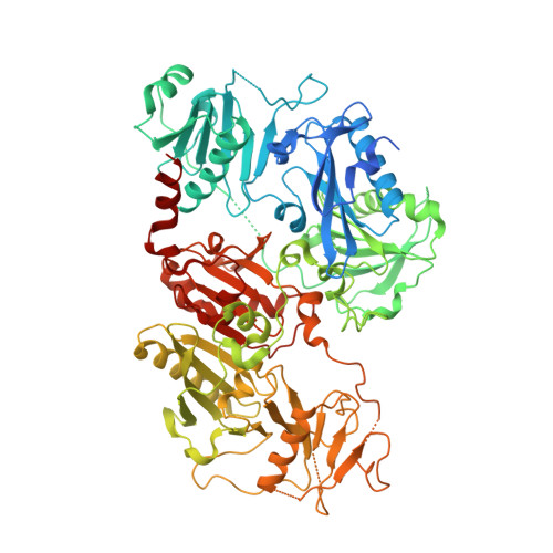
6Q9R, 6Q9Z, 6QBF - PubMed Abstract:
Mutations in the gelsolin protein are responsible for a rare conformational disease known as AGel amyloidosis. Four of these mutations are hosted by the second domain of the protein (G2): D187N/Y, G167R and N184K. The impact of the latter has been so far evaluated only by studies on the isolated G2. Here we report the characterization of full-length gelsolin carrying the N184K mutation and compare the findings with those obtained on the wild type and the other variants. The crystallographic structure of the N184K variant in the Ca 2+ -free conformation shows remarkable similarities with the wild type protein. Only minimal local rearrangements can be observed and the mutant is as efficient as the wild type in severing filamentous actin. However, the thermal stability of the pathological variant is compromised in the Ca 2+ -free conditions. These data suggest that the N to K substitution causes a local disruption of the H-bond network in the core of the G2 domain. Such a subtle rearrangement of the connections does not lead to significant conformational changes but severely affects the stability of the protein.
- Istituto di Biofisica, Consiglio Nazionale delle Ricerche, via Celoria 26, 20133, Milan, Italy. matteo.derosa@cnr.it.
Organizational Affiliation: