6Q6R | pdb_00006q6r

Recognition of different base tetrads by RHAU: X-ray crystal structure of G4 recognition motif bound to the 3-end tetrad of a DNA G-quadruplex


Experimental Data Snapshot

  • Method: X-RAY DIFFRACTION
  • Resolution: 1.50 Å
  • R-Value Free: 
    0.245 (Depositor), 0.247 (DCC) 
  • R-Value Work: 
    0.185 (Depositor), 0.192 (DCC) 
  • R-Value Observed: 
    0.188 (Depositor) 

Starting Model: experimental
View more details

wwPDB Validation 3D Report Full Report

Validation slider image for 6Q6R

This is version 1.4 of the entry. See complete history

Literature

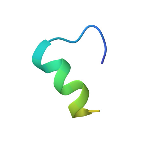
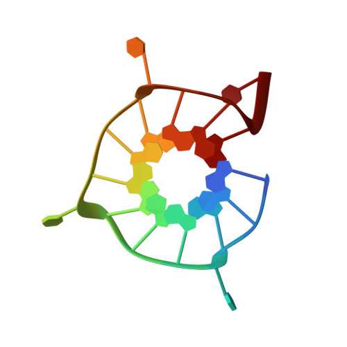
Recognition of different base tetrads by RHAU (DHX36): X-ray crystal structure of the G4 recognition motif bound to the 3'-end tetrad of a DNA G-quadruplex.

Heddi, B.Cheong, V.V.Schmitt, E.Mechulam, Y.Phan, A.T.

(2020) J Struct Biol 209: 107399-107399

  • DOI: https://doi.org/10.1016/j.jsb.2019.10.001
  • Primary Citation Related Structures: 
    6Q6R

  • PubMed Abstract: 

    G-quadruplexes (G4) are secondary structures of nucleic acids that can form in cells and have diverse biological functions. Several biologically important proteins interact with G-quadruplexes, of which RHAU (or DHX36) - a helicase from the DEAH-box superfamily, was shown to bind and unwind G-quadruplexes efficiently. We report a X-ray co-crystal structure at 1.5 Å resolution of an N-terminal fragment of RHAU bound to an exposed tetrad of a parallel-stranded G-quadruplex. The RHAU peptide folds into an L-shaped α-helix, and binds to a G-quadruplex through π-stacking and electrostatic interactions. X-ray crystal structure of our complex identified key amino acid residues important for G-quadruplex-peptide binding interaction at the 3'-end G•G•G•G tetrad. Together with previous solution and crystal structures of RHAU bound to the 5'-end G•G•G•G and G•G•A•T tetrads, our crystal structure highlights the occurrence of a robust G-quadruplex recognition motif within RHAU that can adapt to different accessible tetrads.


  • Organizational Affiliation
    • School of Physical and Mathematical Sciences, Nanyang Technological University, 21 Nanyang Link, Singapore 637371, Singapore; Laboratoire de Biologie et de Pharmacologie Appliquée, CNRS UMR 8113, Ecole Normale Supérieure Paris-Saclay, 61 Avenue du président Wilson, Cachan 94235, France. Electronic address: brahim.heddi@ens-cachan.fr.

Macromolecule Content 

  • Total Structure Weight: 34.66 kDa 
  • Atom Count: 2,228 
  • Modeled Residue Count: 132 
  • Deposited Residue Count: 180 
  • Unique protein chains: 1
  • Unique nucleic acid chains: 1

Macromolecules


Find similar proteins by:|  3D Structure
Entity ID: 2
MoleculeChains  Sequence LengthOrganismDetailsImage
ATP-dependent DNA/RNA helicase DHX36
E, F, G, H
29Homo sapiensMutation(s): 0 
Gene Names: DHX36DDX36KIAA1488MLEL1RHAU
EC: 3.6.4.12 (PDB Primary Data), 3.6.4.13 (PDB Primary Data)
UniProt & NIH Common Fund Data Resources
Find proteins for Q9H2U1 (Homo sapiens)
Explore Q9H2U1 
Go to UniProtKB:  Q9H2U1
PHAROS:  Q9H2U1
GTEx:  ENSG00000174953 
Entity Groups
Sequence Clusters30% Identity50% Identity70% Identity90% Identity95% Identity100% Identity
UniProt GroupQ9H2U1
Sequence Annotations
Expand
Reference Sequence
Find similar nucleic acids by:  Sequence
Entity ID: 1
MoleculeChains LengthOrganismImage
Parallel stranded DNA G-quadruplex
A, B, C, D
16unidentified
Sequence Annotations
Expand
Reference Sequence

Small Molecules

Ligands 1 Unique
IDChains Name / Formula / InChI Key2D Diagram3D Interactions
K

Query on K


Download:Ideal Coordinates CCD File
I [auth A]
J [auth A]
K [auth B]
L [auth B]
M [auth B]
I [auth A],
J [auth A],
K [auth B],
L [auth B],
M [auth B],
N [auth C],
O [auth C],
P [auth C],
Q [auth D],
R [auth D]
POTASSIUM ION
K
NPYPAHLBTDXSSS-UHFFFAOYSA-N

Experimental Data & Validation

Experimental Data

  • Method: X-RAY DIFFRACTION
  • Resolution: 1.50 Å
  • R-Value Free:  0.245 (Depositor), 0.247 (DCC) 
  • R-Value Work:  0.185 (Depositor), 0.192 (DCC) 
  • R-Value Observed: 0.188 (Depositor) 
Space Group: P 1 21 1
Unit Cell:
Length ( Å )Angle ( ˚ )
a = 56.39α = 90
b = 42.25β = 99.3
c = 61.404γ = 90
Software Package:
Software NamePurpose
REFMACrefinement
XDSdata reduction
XDSdata scaling
PHASERphasing

Structure Validation

View Full Validation Report


Entry History 

& Funding Information

Deposition Data


Funding OrganizationLocationGrant Number
National Research Foundation (Singapore)SingaporeNRF-NRFI2017-09
Ministry of Education (Singapore)SingaporeMOE2012-T3-1-001

Revision History  (Full details and data files)

  • Version 1.0: 2019-10-16
    Type: Initial release
  • Version 1.1: 2019-11-27
    Changes: Database references
  • Version 1.2: 2020-01-22
    Changes: Database references
  • Version 1.3: 2022-07-13
    Changes: Database references, Derived calculations, Structure summary
  • Version 1.4: 2024-01-24
    Changes: Data collection, Refinement description