Structural and functional characterization of three Type B and C chloramphenicol acetyltransferases from Vibrio species.
Alcala, A., Ramirez, G., Solis, A., Kim, Y., Tan, K., Luna, O., Nguyen, K., Vazquez, D., Ward, M., Zhou, M., Mulligan, R., Maltseva, N., Kuhn, M.L.(2020) Protein Sci 29: 695-710
- PubMed: 31762145 Search on PubMedSearch on PubMed Central
- DOI: https://doi.org/10.1002/pro.3793
- Primary Citation Related Structures:
3EEV, 6PUB - PubMed Abstract:
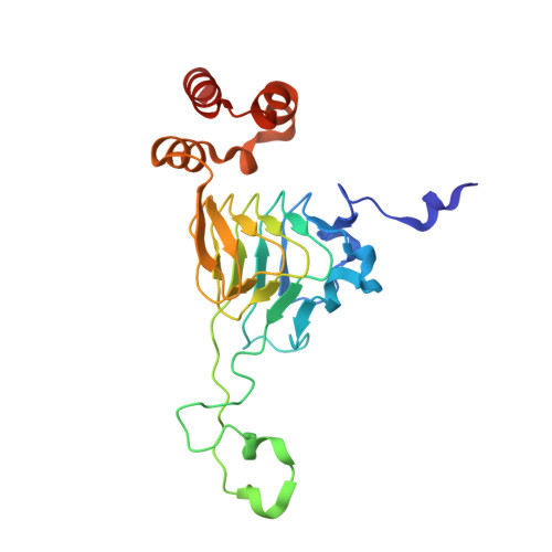
Chloramphenicol acetyltransferases (CATs) were among the first antibiotic resistance enzymes identified and have long been studied as model enzymes for examining plasmid-mediated antibiotic resistance. These enzymes acetylate the antibiotic chloramphenicol, which renders it incapable of inhibiting bacterial protein synthesis. CATs can be classified into different types: Type A CATs are known to be important for antibiotic resistance to chloramphenicol and fusidic acid. Type B CATs are often called xenobiotic acetyltransferases and adopt a similar structural fold to streptogramin acetyltransferases, which are known to be critical for streptogramin antibiotic resistance. Type C CATs have recently been identified and can also acetylate chloramphenicol, but their roles in antibiotic resistance are largely unknown. Here, we structurally and kinetically characterized three Vibrio CAT proteins from a nonpathogenic species (Aliivibrio fisheri) and two important human pathogens (Vibrio cholerae and Vibrio vulnificus). We found all three proteins, including one in a superintegron (V. cholerae), acetylated chloramphenicol, but did not acetylate aminoglycosides or dalfopristin. We also determined the 3D crystal structures of these CATs alone and in complex with crystal violet and taurocholate. These compounds are known inhibitors of Type A CATs, but have not been explored in Type B and Type C CATs. Based on sequence, structure, and kinetic analysis, we concluded that the V. cholerae and V. vulnificus CATs belong to the Type B class and the A. fisheri CAT belongs to the Type C class. Ultimately, our results provide a framework for studying the evolution of antibiotic resistance gene acquisition and chloramphenicol acetylation in Vibrio and other species.
- San Francisco State University, Department of Chemistry and Biochemistry, San Francisco, California.
Organizational Affiliation: