6PPM | pdb_00006ppm

Ancestral Caspase 6

  • Classification: APOPTOSIS
  • Organism(s): Homo sapiens
  • Expression System: Escherichia coli
  • Mutation(s): No 

  • Deposited: 2019-07-08 Released: 2019-11-13 
  • Deposition Author(s): Clark, A.C.
  • Funding Organization(s): National Institutes of Health/National Institute of General Medical Sciences (NIH/NIGMS)

Experimental Data Snapshot

  • Method: X-RAY DIFFRACTION
  • Resolution: 2.61 Å
  • R-Value Free: 
    0.251 (Depositor), 0.251 (DCC) 
  • R-Value Work: 
    0.177 (Depositor), 0.180 (DCC) 
  • R-Value Observed: 
    0.182 (Depositor) 

Starting Model: experimental
View more details

wwPDB Validation 3D Report Full Report

Validation slider image for 6PPM

This is version 1.3 of the entry. See complete history

Literature

Resurrection of ancestral effector caspases identifies novel networks for evolution of substrate specificity.

Grinshpon, R.D.Shrestha, S.Titus-McQuillan, J.Hamilton, P.T.Swartz, P.D.Clark, A.C.

(2019) Biochem J 476: 3475-3492

  • DOI: https://doi.org/10.1042/BCJ20190625
  • Primary Citation Related Structures: 
    6PDQ, 6PPM

  • PubMed Abstract: 

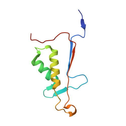
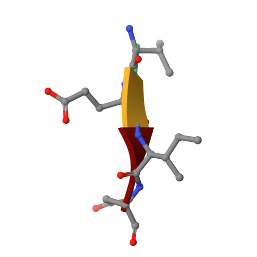
    Apoptotic caspases evolved with metazoans more than 950 million years ago (MYA), and a series of gene duplications resulted in two subfamilies consisting of initiator and effector caspases. The effector caspase genes (caspases-3, -6, and -7) were subsequently fixed into the Chordata phylum more than 650 MYA when the gene for a common ancestor (CA) duplicated, and the three effector caspases have persisted throughout mammalian evolution. All caspases prefer an aspartate residue at the P1 position of substrates, so each caspase evolved discrete cellular roles through changes in substrate recognition at the P4 position combined with allosteric regulation. We examined the evolution of substrate specificity in caspase-6, which prefers valine at the P4 residue, compared with caspases-3 and -7, which prefer aspartate, by reconstructing the CA of effector caspases (AncCP-Ef1) and the CA of caspase-6 (AncCP-6An). We show that AncCP-Ef1 is a promiscuous enzyme with little distinction between Asp, Val, or Leu at P4. The specificity of caspase-6 was defined early in its evolution, where AncCP-6An demonstrates a preference for Val over Asp at P4. Structures of AncCP-Ef1 and of AncCP-6An show a network of charged amino acids near the S4 pocket that, when combined with repositioning a flexible active site loop, resulted in a more hydrophobic binding pocket in AncCP-6An. The ancestral protein reconstructions show that the caspase-hemoglobinase fold has been conserved for over 650 million years and that only three substitutions in the scaffold are necessary to shift substrate selection toward Val over Asp.


  • Organizational Affiliation
    • Department of Biology, University of Texas at Arlington, Arlington, TX 76019, U.S.A.

Macromolecule Content 

  • Total Structure Weight: 115.59 kDa 
  • Atom Count: 7,697 
  • Modeled Residue Count: 974 
  • Deposited Residue Count: 1,000 
  • Unique protein chains: 3

Macromolecules

Find similar proteins by:|  3D Structure
Entity ID: 1
MoleculeChains  Sequence LengthOrganismDetailsImage
Ancestral Caspase-6 Large SubunitA,
D [auth G],
G [auth C],
I [auth J]
151Homo sapiensMutation(s): 0 
Gene Names: CASP6MCH2
EC: 3.4.22.59
Find similar proteins by:|  3D Structure
Entity ID: 2
MoleculeChains  Sequence LengthOrganismDetailsImage
Ancestral Caspase-6 small subunitB,
E [auth H],
H [auth D],
J [auth K]
95Homo sapiensMutation(s): 0 
Gene Names: CASP6MCH2
EC: 3.4.22.59
Find similar proteins by:  Sequence   |   3D Structure  
Entity ID: 3
MoleculeChains  Sequence LengthOrganismDetailsImage
VAL-GLU-ILE-ASP InhibitorC [auth E],
F [auth I],
K [auth L],
L [auth F]
4Homo sapiensMutation(s): 0 

Experimental Data & Validation

Experimental Data

  • Method: X-RAY DIFFRACTION
  • Resolution: 2.61 Å
  • R-Value Free:  0.251 (Depositor), 0.251 (DCC) 
  • R-Value Work:  0.177 (Depositor), 0.180 (DCC) 
  • R-Value Observed: 0.182 (Depositor) 
Space Group: P 21 21 21
Unit Cell:
Length ( Å )Angle ( ˚ )
a = 84.314α = 90
b = 88.542β = 90
c = 141.026γ = 90
Software Package:
Software NamePurpose
PHENIXrefinement
HKL-2000data reduction
HKL-2000data scaling
PHENIXphasing

Structure Validation

View Full Validation Report


Entry History 

& Funding Information

Deposition Data

  • Released Date: 2019-11-13 
  • Deposition Author(s): Clark, A.C.

Funding OrganizationLocationGrant Number
National Institutes of Health/National Institute of General Medical Sciences (NIH/NIGMS)United StatesGM127654

Revision History  (Full details and data files)

  • Version 1.0: 2019-11-13
    Type: Initial release
  • Version 1.1: 2019-12-04
    Changes: Database references
  • Version 1.2: 2020-01-01
    Changes: Author supporting evidence
  • Version 1.3: 2023-10-11
    Changes: Advisory, Data collection, Database references, Refinement description