Crystal Structure Of Photorespiratory Alanine:Glyoxylate Aminotransferase 1 (AGT1) FromArabidopsis thaliana.
Liepman, A.H., Vijayalakshmi, J., Peisach, D., Hulsebus, B., Olsen, L.J., Saper, M.A.(2019) Front Plant Sci 10: 1229-1229
- PubMed: 31681359 Search on PubMedSearch on PubMed Central
- DOI: https://doi.org/10.3389/fpls.2019.01229
- Primary Citation Related Structures:
6PK1, 6PK3 - PubMed Abstract:
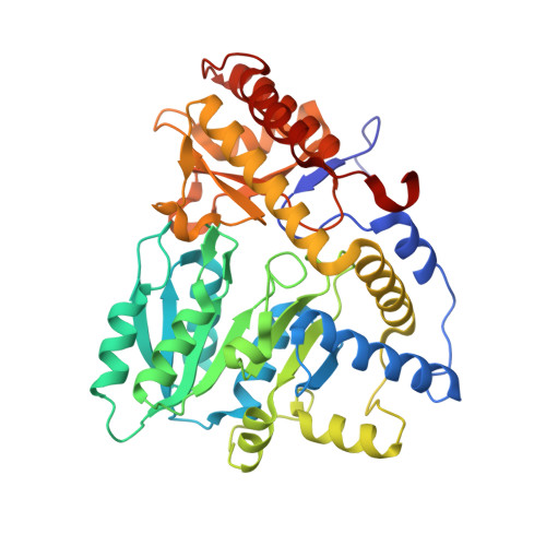
Photorespiration is an energetically costly metabolic pathway for the recycling of phosphoglycolate produced by the oxygenase activity of ribulose-1,5-bisphosphate carboxylase/oxygenase (RUBISCO) to phosphoglycerate. Arabidopsis alanine:glyoxylate aminotransferase 1 (AGT1) is a peroxisomal aminotransferase with a central role in photorespiration. This enzyme catalyzes various aminotransferase reactions, including serine:glyoxylate, alanine:glyoxylate, and asparagine:glyoxylate transaminations. To better understand structural features that govern the specificity of this enzyme, its crystal structures in the native form (2.2-Å resolution) and in the presence of l-serine (2.1-Å resolution) were solved. The structures confirm that this enzyme is dimeric, in agreement with studies of the active enzyme in solution. In the crystal, another dimer related by noncrystallographic symmetry makes close interactions to form a tetramer mediated in part by an extra carboxyl-terminal helix conserved in plant homologs of AGT1. Pyridoxal 5'-phosphate (PLP) is bound at the active site but is not held in place by covalent interactions. Residues Tyr35' and Arg36', entering the active site from the other subunits in the dimer, mediate interactions between AGT and l-serine when used as a substrate. In comparison, AGT1 from humans and AGT1 from Anabaena lack these two residues and instead position a tyrosine ring into the binding site, which accounts for their preference for l-alanine instead of l-serine. The structure also rationalizes the phenotype of the sat mutant, Pro251 to Leu, which likely affects the dimer interface near the catalytic site. This structural model of AGT1 provides valuable new information about this protein that may enable improvements to the efficiency of photorespiration.
- Biology Department, Eastern Michigan University, Ypsilanti, MI, United States.
Organizational Affiliation: