6PEE

InvG secretin domain beta-barrel from Salmonella SPI-1 injectisome NC-base


Experimental Data Snapshot

  • Method: ELECTRON MICROSCOPY
  • Resolution: 3.42 Å
  • Aggregation State: PARTICLE 
  • Reconstruction Method: SINGLE PARTICLE 

wwPDB Validation   3D Report Full Report


This is version 1.3 of the entry. See complete history


Literature

T3S injectisome needle complex structures in four distinct states reveal the basis of membrane coupling and assembly.

Hu, J.Worrall, L.J.Vuckovic, M.Hong, C.Deng, W.Atkinson, C.E.Brett Finlay, B.Yu, Z.Strynadka, N.C.J.

(2019) Nat Microbiol 4: 2010-2019

  • DOI: https://doi.org/10.1038/s41564-019-0545-z
  • Primary Citation of Related Structures:  
    6PEE, 6PEM, 6PEP, 6Q14, 6Q15, 6Q16

  • PubMed Abstract: 

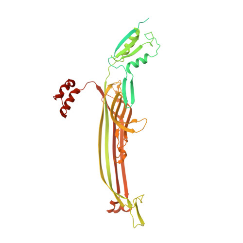
    The bacterial injectisome is a syringe-shaped macromolecular nanomachine utilized by many pathogenic Gram-negative bacteria, including the causative agents of plague, typhoid fever, whooping cough, sexually transmitted infections and major nosocomial infections. Bacterial proteins destined for self-assembly and host-cell targeting are translocated by the injectisome in a process known as type III secretion (T3S). The core structure is the ~4 MDa needle complex (NC), built on a foundation of three highly oligomerized ring-forming proteins that create a hollow scaffold spanning the bacterial inner membrane (IM) (24-mer ring-forming proteins PrgH and PrgK in the Salmonella enterica serovar Typhimurium Salmonella pathogenicity island 1 (SPI-1) type III secretion system (T3SS)) and outer membrane (OM) (15-mer InvG, a member of the broadly conserved secretin pore family). An internalized helical needle projects from the NC and bacterium, ultimately forming a continuous passage to the host, for delivery of virulence effectors. Here, we have captured snapshots of the entire prototypical SPI-1 NC in four distinct needle assembly states, including near-atomic resolution, and local reconstructions in the absence and presence of the needle. These structures reveal the precise localization and molecular interactions of the internalized SpaPQR 'export apparatus' complex, which is intimately encapsulated and stabilized within the IM rings in the manner of a nanodisc, and to which the PrgJ rod directly binds and functions as an initiator and anchor of needle polymerization. We also describe the molecular details of the extensive and continuous coupling interface between the OM secretin and IM rings, which is remarkably facilitated by a localized 16-mer stoichiometry in the periplasmic-most coupling domain of the otherwise 15-mer InvG oligomer.


  • Organizational Affiliation

    Department of Biochemistry and Molecular Biology, University of British Columbia, Vancouver, British Columbia, Canada.


Macromolecules
Find similar proteins by:  (by identity cutoff)  |  3D Structure
Entity ID: 1
MoleculeChains Sequence LengthOrganismDetailsImage
Protein InvG562Salmonella enterica subsp. enterica serovar Typhimurium str. LT2Mutation(s): 0 
Membrane Entity: Yes 
UniProt
Find proteins for P35672 (Salmonella typhimurium (strain LT2 / SGSC1412 / ATCC 700720))
Explore P35672 
Go to UniProtKB:  P35672
Entity Groups  
Sequence Clusters30% Identity50% Identity70% Identity90% Identity95% Identity100% Identity
UniProt GroupP35672
Sequence Annotations
Expand
  • Reference Sequence
Small Molecules
Ligands 1 Unique
IDChains Name / Formula / InChI Key2D Diagram3D Interactions
LDA
Query on LDA

Download Ideal Coordinates CCD File 
AA [auth D]
AB [auth N]
BA [auth F]
BB [auth N]
CA [auth F]
AA [auth D],
AB [auth N],
BA [auth F],
BB [auth N],
CA [auth F],
CB [auth O],
DA [auth F],
DB [auth O],
EA [auth G],
EB [auth O],
FA [auth G],
FB [auth P],
GA [auth G],
GB [auth P],
HA [auth H],
HB [auth P],
IA [auth H],
JA [auth H],
KA [auth I],
LA [auth I],
MA [auth I],
NA [auth J],
OA [auth J],
P [auth A],
PA [auth J],
Q [auth A],
QA [auth K],
R [auth A],
RA [auth K],
S [auth B],
SA [auth K],
T [auth B],
TA [auth L],
U [auth B],
UA [auth L],
V [auth C],
VA [auth L],
W [auth C],
WA [auth M],
X [auth C],
XA [auth M],
Y [auth D],
YA [auth M],
Z [auth D],
ZA [auth N]
LAURYL DIMETHYLAMINE-N-OXIDE
C14 H31 N O
SYELZBGXAIXKHU-UHFFFAOYSA-N
Experimental Data & Validation

Experimental Data

  • Method: ELECTRON MICROSCOPY
  • Resolution: 3.42 Å
  • Aggregation State: PARTICLE 
  • Reconstruction Method: SINGLE PARTICLE 

Structure Validation

View Full Validation Report


Entry History & Funding Information

Deposition Data


Funding OrganizationLocationGrant Number
Canadian Institutes of Health Research (CIHR)Canada--

Revision History  (Full details and data files)

  • Version 1.0: 2019-10-23
    Type: Initial release
  • Version 1.1: 2019-11-06
    Changes: Data collection, Database references
  • Version 1.2: 2020-01-08
    Changes: Author supporting evidence
  • Version 1.3: 2024-03-13
    Changes: Data collection, Database references