6PD4

Crystal Structure of Hendra Virus Attachment G Glycoprotein


Experimental Data Snapshot

  • Method: X-RAY DIFFRACTION
  • Resolution: 2.20 Å
  • R-Value Free: 0.207 
  • R-Value Work: 0.167 
  • R-Value Observed: 0.169 

wwPDB Validation   3D Report Full Report


Ligand Structure Quality Assessment 


This is version 2.0 of the entry. See complete history


Literature

New insights into the Hendra virus attachment and entry process from structures of the virus G glycoprotein and its complex with Ephrin-B2.

Xu, K.Chan, Y.P.Rajashankar, K.R.Khetawat, D.Yan, L.Kolev, M.V.Broder, C.C.Nikolov, D.B.

(2012) PLoS One 7: e48742

  • DOI: https://doi.org/10.1371/journal.pone.0048742
  • Primary Citation of Related Structures:  
    6PD4, 6PDL

  • PubMed Abstract: 

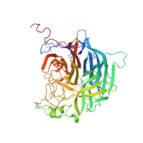
    Hendra virus and Nipah virus, comprising the genus Henipavirus, are recently emerged, highly pathogenic and often lethal zoonotic agents against which there are no approved therapeutics. Two surface glycoproteins, the attachment (G) and fusion (F), mediate host cell entry. The crystal structures of the Hendra G glycoprotein alone and in complex with the ephrin-B2 receptor reveal that henipavirus uses Tryptophan 122 on ephrin-B2/B3 as a "latch" to facilitate the G-receptor association. Structural-based mutagenesis of residues in the Hendra G glycoprotein at the receptor binding interface document their importance for viral attachments and entry, and suggest that the stability of the Hendra-G-ephrin attachment complex does not strongly correlate with the efficiency of viral entry. In addition, our data indicates that conformational rearrangements of the G glycoprotein head domain upon receptor binding may be the trigger leading to the activation of the viral F fusion glycoprotein during virus infection.


  • Organizational Affiliation

    Structural Biology Program, Memorial Sloan-Kettering Cancer Center, New York, New York, United States of America.


Macromolecules
Find similar proteins by:  (by identity cutoff)  |  3D Structure
Entity ID: 1
MoleculeChains Sequence LengthOrganismDetailsImage
Attachment glycoprotein
A, B
441Henipavirus hendraenseMutation(s): 0 
UniProt
Find proteins for O89343 (Hendra virus (isolate Horse/Autralia/Hendra/1994))
Explore O89343 
Go to UniProtKB:  O89343
Entity Groups  
Sequence Clusters30% Identity50% Identity70% Identity90% Identity95% Identity100% Identity
UniProt GroupO89343
Sequence Annotations
Expand
  • Reference Sequence
Oligosaccharides

Help

Entity ID: 2
MoleculeChains Length2D Diagram Glycosylation3D Interactions
2-acetamido-2-deoxy-beta-D-glucopyranose-(1-4)-2-acetamido-2-deoxy-beta-D-glucopyranose
C, D, F, G
2N-Glycosylation
Glycosylation Resources
GlyTouCan:  G42666HT
GlyCosmos:  G42666HT
GlyGen:  G42666HT
Entity ID: 3
MoleculeChains Length2D Diagram Glycosylation3D Interactions
alpha-L-fucopyranose-(1-3)-[2-acetamido-2-deoxy-beta-D-glucopyranose-(1-4)][alpha-L-fucopyranose-(1-6)]2-acetamido-2-deoxy-beta-D-glucopyranose
E
4N-Glycosylation
Glycosylation Resources
GlyTouCan:  G63564LA
GlyCosmos:  G63564LA
Entity ID: 4
MoleculeChains Length2D Diagram Glycosylation3D Interactions
beta-D-mannopyranose-(1-6)-beta-D-mannopyranose-(1-4)-2-acetamido-2-deoxy-beta-D-glucopyranose-(1-4)-[alpha-L-fucopyranose-(1-3)][alpha-L-fucopyranose-(1-6)]2-acetamido-2-deoxy-beta-D-glucopyranose
H
6N-Glycosylation
Glycosylation Resources
GlyTouCan:  G57322IK
GlyCosmos:  G57322IK
GlyGen:  G57322IK
Small Molecules
Ligands 2 Unique
IDChains Name / Formula / InChI Key2D Diagram3D Interactions
NAG
Query on NAG

Download Ideal Coordinates CCD File 
I [auth A],
Q [auth B]
2-acetamido-2-deoxy-beta-D-glucopyranose
C8 H15 N O6
OVRNDRQMDRJTHS-FMDGEEDCSA-N
SO4
Query on SO4

Download Ideal Coordinates CCD File 
J [auth A]
K [auth A]
L [auth A]
M [auth A]
N [auth A]
J [auth A],
K [auth A],
L [auth A],
M [auth A],
N [auth A],
O [auth A],
P [auth A],
R [auth B],
S [auth B],
T [auth B],
U [auth B],
V [auth B],
W [auth B]
SULFATE ION
O4 S
QAOWNCQODCNURD-UHFFFAOYSA-L
Experimental Data & Validation

Experimental Data

  • Method: X-RAY DIFFRACTION
  • Resolution: 2.20 Å
  • R-Value Free: 0.207 
  • R-Value Work: 0.167 
  • R-Value Observed: 0.169 
  • Space Group: P 1 21 1
Unit Cell:
Length ( Å )Angle ( ˚ )
a = 66.152α = 90
b = 73.244β = 91.85
c = 109.132γ = 90
Software Package:
Software NamePurpose
PHENIXrefinement
HKL-2000data scaling
PDB_EXTRACTdata extraction
HKL-2000data reduction
PHASERphasing

Structure Validation

View Full Validation Report


Ligand Structure Quality Assessment 


Entry History & Funding Information

Deposition Data


Funding OrganizationLocationGrant Number
National Institutes of Health/National Human Genome Research Institute (NIH/NHGRI)United StatesNS38486
National Institutes of Health/National Human Genome Research Institute (NIH/NHGRI)United StatesAI057168
National Institutes of Health/National Human Genome Research Institute (NIH/NHGRI)United StatesAI054715

Revision History  (Full details and data files)

  • Version 1.0: 2019-11-27
    Type: Initial release
  • Version 1.1: 2019-12-18
    Changes: Author supporting evidence
  • Version 2.0: 2020-07-29
    Type: Remediation
    Reason: Carbohydrate remediation
    Changes: Advisory, Atomic model, Data collection, Derived calculations, Structure summary