6P67 | pdb_00006p67

Crystal Structure of a Complex of human IL-7Ralpha with an anti-IL-7Ralpha 2B8 Fab


Experimental Data Snapshot

  • Method: X-RAY DIFFRACTION
  • Resolution: 2.90 Å
  • R-Value Free: 
    0.266 (Depositor) 
  • R-Value Work: 
    0.249 (Depositor) 
  • R-Value Observed: 
    0.249 (Depositor) 

Starting Models: experimental
View more details

wwPDB Validation 3D Report Full Report

Validation slider image for 6P67

Ligand Structure Quality Assessment 


This is version 2.3 of the entry. See complete history

Literature

New anti-IL-7R alpha monoclonal antibodies show efficacy against T cell acute lymphoblastic leukemia in pre-clinical models.

Hixon, J.A.Andrews, C.Kashi, L.Kohnhorst, C.L.Senkevitch, E.Czarra, K.Barata, J.T.Li, W.Schneider, J.P.Walsh, S.T.R.Durum, S.K.

(2020) Leukemia 34: 35-49

  • DOI: https://doi.org/10.1038/s41375-019-0531-8
  • Primary Citation Related Structures: 
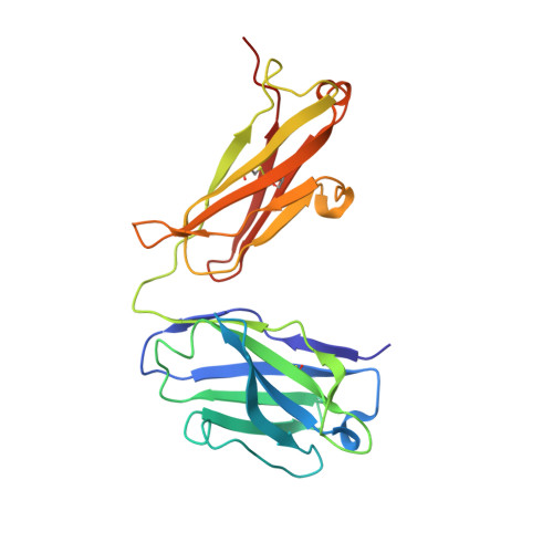
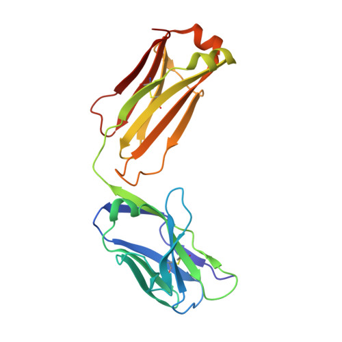
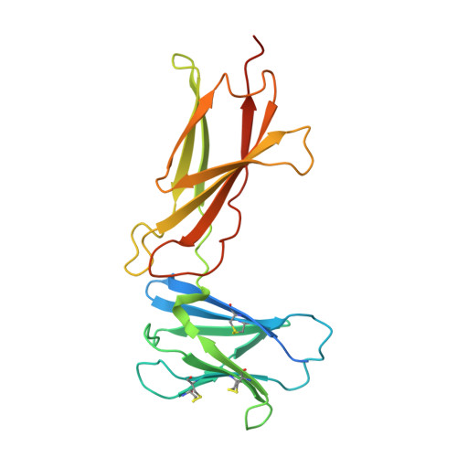
    6P4Y, 6P50, 6P67

  • PubMed Abstract: 

    Pediatric T cell acute lymphoblastic leukemia (T-ALL) cells frequently contain mutations in the interleukin-7 (IL-7) receptor pathway or respond to IL-7 itself. To target the IL-7 receptor on T-ALL cells, murine monoclonal antibodies (MAbs) were developed against the human IL-7Rα chain and chimerized with human IgG1 constant regions. Crystal structures demonstrate that the two MAbs bound different IL-7Rα epitopes. The MAbs mediated antibody-dependent cell-mediated cytotoxicity (ADCC) against patient-derived xenograft (PDX) T-ALL cells, which was improved by combining two MAbs. In vivo, the MAbs showed therapeutic efficacy via ADCC-dependent and independent mechanisms in minimal residual and established disease. PDX T-ALL cells that relapsed following a course of chemotherapy displayed elevated IL-7Rα, and MAb treatment is effective against relapsing disease, suggesting the use of anti-IL7Rα MAbs in relapsed T-ALL patients or patients that do not respond to chemotherapy.


  • Organizational Affiliation
    • Cytokines and Immunity Section, Cancer and Inflammation Program (CIP), National Cancer Institute (NCI), National Institutes of Health (NIH), Frederick, MD, USA.

Macromolecule Content 

  • Total Structure Weight: 296.91 kDa 
  • Atom Count: 19,325 
  • Modeled Residue Count: 2,512 
  • Deposited Residue Count: 2,648 
  • Unique protein chains: 3

Macromolecules

Find similar proteins by:|  3D Structure
Entity ID: 1
MoleculeChains  Sequence LengthOrganismDetailsImage
Anti-IL-7R 2B8 Fab heavy chain
A, C, E, H
225Mus musculusMutation(s): 0 
Find similar proteins by:|  3D Structure
Entity ID: 2
MoleculeChains  Sequence LengthOrganismDetailsImage
anti-IL-7R 2B8 Fab light chain
B, D, F, L
214Mus musculusMutation(s): 0 
Find similar proteins by:|  3D Structure
Entity ID: 3
MoleculeChains  Sequence LengthOrganismDetailsImage
Interleukin-7 receptor subunit alpha
G, I, J, K
223Homo sapiensMutation(s): 0 
Gene Names: IL7R
UniProt & NIH Common Fund Data Resources
Find proteins for P16871 (Homo sapiens)
Explore P16871 
Go to UniProtKB:  P16871
PHAROS:  P16871
GTEx:  ENSG00000168685 
Entity Groups
Sequence Clusters30% Identity50% Identity70% Identity90% Identity95% Identity100% Identity
UniProt GroupP16871
Glycosylation
Glycosylation Sites: 1Go to GlyGen: P16871-1
Sequence Annotations
Expand
Reference Sequence

Oligosaccharides

Help  
Entity ID: 4
MoleculeChains Length2D Diagram GlycosylationD Interactions
beta-D-mannopyranose-(1-4)-2-acetamido-2-deoxy-beta-D-glucopyranose-(1-4)-2-acetamido-2-deoxy-beta-D-glucopyranose
M, N
3N-Glycosylation
Glycosylation Resources
GlyTouCan: G15407YE
GlyCosmos: G15407YE
GlyGen: G15407YE
Entity ID: 5
MoleculeChains Length2D Diagram GlycosylationD Interactions
2-acetamido-2-deoxy-beta-D-glucopyranose-(1-4)-2-acetamido-2-deoxy-beta-D-glucopyranose
O
2N-Glycosylation
Glycosylation Resources
GlyTouCan: G42666HT
GlyCosmos: G42666HT
GlyGen: G42666HT

Experimental Data & Validation

Experimental Data

  • Method: X-RAY DIFFRACTION
  • Resolution: 2.90 Å
  • R-Value Free:  0.266 (Depositor) 
  • R-Value Work:  0.249 (Depositor) 
  • R-Value Observed: 0.249 (Depositor) 
Space Group: P 1 21 1
Unit Cell:
Length ( Å )Angle ( ˚ )
a = 84.9α = 90
b = 219.68β = 104.328
c = 90.35γ = 90
Software Package:
Software NamePurpose
phenix.refinerefinement
PHENIXrefinement
XDSdata scaling
PDB_EXTRACTdata extraction
XDSdata reduction
PHASERphasing

Structure Validation

View Full Validation Report


Ligand Structure Quality Assessment 


Entry History 

& Funding Information

Deposition Data


Funding OrganizationLocationGrant Number
National Institutes of Health/National Cancer Institute (NIH/NCI)United States--

Revision History  (Full details and data files)

  • Version 1.0: 2019-09-04
    Type: Initial release
  • Version 1.1: 2019-12-04
    Changes: Author supporting evidence, Data collection
  • Version 1.2: 2020-01-01
    Changes: Database references
  • Version 2.0: 2020-07-29
    Type: Remediation
    Reason: Carbohydrate remediation
    Changes: Advisory, Atomic model, Data collection, Derived calculations, Source and taxonomy, Structure summary
  • Version 2.1: 2023-10-11
    Changes: Data collection, Database references, Refinement description, Structure summary
  • Version 2.2: 2024-03-13
    Changes: Source and taxonomy
  • Version 2.3: 2024-10-23
    Changes: Structure summary