6OW7 | pdb_00006ow7

X-ray Structure of Polypeptide Deformylase with a Piperazic Acid


Experimental Data Snapshot

  • Method: X-RAY DIFFRACTION
  • Resolution: 1.45 Å
  • R-Value Free: 
    0.218 (Depositor), 0.238 (DCC) 
  • R-Value Work: 
    0.206 (Depositor) 
  • R-Value Observed: 
    0.206 (Depositor) 

wwPDB Validation 3D Report Full Report

Validation slider image for 6OW7

Ligand Structure Quality Assessment 


This is version 1.2 of the entry. See complete history

Literature

Discovery of piperazic acid peptide deformylase inhibitors with in vivo activity for respiratory tract and skin infections.

Spletstoser, J.T.Dreabit, J.Knox, A.N.Benowitz, A.Campobasso, N.Ward, P.Cui, G.Lewandowski, T.McCloskey, L.Aubart, K.M.

(2019) Bioorg Med Chem Lett 29: 2410-2414

  • DOI: https://doi.org/10.1016/j.bmcl.2019.05.028
  • Primary Citation Related Structures: 
    6OW2, 6OW7

  • PubMed Abstract: 

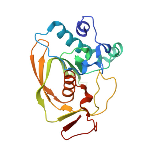
    The discovery of a novel series of peptide deformylase inhibitors incorporating a piperazic acid amino acid found in nature is described. These compounds demonstrated potent in vitro enzymatic potency and antimicrobial activity. Crystal structure analysis revealed the piperazic acid optimized a key contact with the PDF protein that accounted for the increased enzymatic potency of these compounds. We describe lead optimization of the P3' region of the series that resulted in a compound with good potency against three target organisms. One molecule showed in vivo efficacy in a rat respiratory infection model but ultimately did not meet candidate progression criteria.


  • Organizational Affiliation
    • GlaxoSmithKline, 1250 S. Collegeville Rd., Collegeville, PA 19426, USA. Electronic address: jared.t.spletstoser@gsk.com.

Macromolecule Content 

  • Total Structure Weight: 46.3 kDa 
  • Atom Count: 3,310 
  • Modeled Residue Count: 387 
  • Deposited Residue Count: 404 
  • Unique protein chains: 1

Macromolecules

Find similar proteins by:|  3D Structure
Entity ID: 1
MoleculeChains  Sequence LengthOrganismDetailsImage
Peptide deformylaseA [auth P],
B [auth Q]
202Streptococcus pneumoniaeMutation(s): 0 
Gene Names: 
EC: 3.5.1.88
UniProt
Find proteins for Q8DP79 (Streptococcus pneumoniae (strain ATCC BAA-255 / R6))
Explore Q8DP79 
Go to UniProtKB:  Q8DP79
Entity Groups
Sequence Clusters30% Identity50% Identity70% Identity90% Identity95% Identity100% Identity
UniProt GroupQ8DP79
Sequence Annotations
Expand
Reference Sequence

Small Molecules

Ligands 3 Unique
IDChains Name / Formula / InChI Key2D Diagram3D Interactions
N9Y
(Subject of Investigation/LOI)

Query on N9Y


Download:Ideal Coordinates CCD File
E [auth P],
H [auth Q]
(3S)-2-{(2R)-2-(cyclopentylmethyl)-3-[formyl(hydroxy)amino]propanoyl}-N-(pyridin-2-yl)hexahydropyridazine-3-carboxamide
C20 H29 N5 O4
IGYIJLMSSQSKHF-SJORKVTESA-N
SO4

Query on SO4


Download:Ideal Coordinates CCD File
D [auth P],
G [auth Q]
SULFATE ION
O4 S
QAOWNCQODCNURD-UHFFFAOYSA-L
NI

Query on NI


Download:Ideal Coordinates CCD File
C [auth P],
F [auth Q]
NICKEL (II) ION
Ni
VEQPNABPJHWNSG-UHFFFAOYSA-N

Experimental Data & Validation

Experimental Data

  • Method: X-RAY DIFFRACTION
  • Resolution: 1.45 Å
  • R-Value Free:  0.218 (Depositor), 0.238 (DCC) 
  • R-Value Work:  0.206 (Depositor) 
  • R-Value Observed: 0.206 (Depositor) 
Space Group: P 43
Unit Cell:
Length ( Å )Angle ( ˚ )
a = 69.229α = 90
b = 69.229β = 90
c = 91.879γ = 90
Software Package:
Software NamePurpose
PHENIXrefinement
HKL-2000data reduction

Structure Validation

View Full Validation Report


Ligand Structure Quality Assessment 


Entry History 

Deposition Data

Revision History  (Full details and data files)

  • Version 1.0: 2019-06-26
    Type: Initial release
  • Version 1.1: 2019-08-21
    Changes: Data collection, Database references
  • Version 1.2: 2024-03-13
    Changes: Data collection, Database references, Derived calculations