Calmodulin disrupts plasma membrane localization of farnesylated KRAS4b by sequestering its lipid moiety.
Grant, B.M.M., Enomoto, M., Back, S.I., Lee, K.Y., Gebregiworgis, T., Ishiyama, N., Ikura, M., Marshall, C.B.(2020) Sci Signal 13
- PubMed: 32234958 Search on PubMed
- DOI: https://doi.org/10.1126/scisignal.aaz0344
- Primary Citation Related Structures:
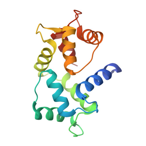
6OS4 - PubMed Abstract:
KRAS4b is a small guanosine triphosphatase (GTPase) protein that regulates several signal transduction pathways that underlie cell proliferation, differentiation, and survival. KRAS4b function requires prenylation of its C terminus and recruitment to the plasma membrane, where KRAS4b activates effector proteins including the RAF family of kinases. The Ca 2+ -sensing protein calmodulin (CaM) has been suggested to regulate the localization of KRAS4b through direct, Ca 2+ -dependent interaction, but how CaM and KRAS4b functionally interact is controversial. Here, we determined a crystal structure, which was supported by solution nuclear magnetic resonance (NMR), that revealed the sequestration of the prenyl moiety of KRAS4b in the hydrophobic pocket of the C-terminal lobe of Ca 2+ -bound CaM. Our engineered fluorescence resonance energy transfer (FRET)-based biosensor probes (CaMeRAS) showed that, upon stimulation of Ca 2+ influx by extracellular ligands, KRAS4b reversibly translocated in a Ca 2+ -CaM-dependent manner from the plasma membrane to the cytoplasm in live HeLa and HEK293 cells. These results reveal a mechanism underlying the inhibition of KRAS4b activity by Ca 2+ signaling pathways.
- Princess Margaret Cancer Center, University Health Network, Toronto, Ontario M5G 1L7, Canada.
Organizational Affiliation: