6OO4

Cryo-EM structure of the C2-symmetric TRPV2/RTx complex in amphipol resolved to 3.3 A


Experimental Data Snapshot

  • Method: ELECTRON MICROSCOPY
  • Resolution: 3.30 Å
  • Aggregation State: PARTICLE 
  • Reconstruction Method: SINGLE PARTICLE 

wwPDB Validation   3D Report Full Report


This is version 1.1 of the entry. See complete history


Literature

Symmetry transitions during gating of the TRPV2 ion channel in lipid membranes.

Zubcevic, L.Hsu, A.L.Borgnia, M.J.Lee, S.Y.

(2019) Elife 8

  • DOI: https://doi.org/10.7554/eLife.45779
  • Primary Citation of Related Structures:  
    6OO3, 6OO4, 6OO5, 6OO7

  • PubMed Abstract: 

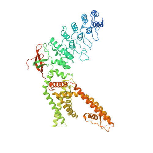
    The Transient Receptor Potential Vanilloid 2 (TRPV2) channel is a member of the temperature-sensing thermoTRPV family. Recent advances in cryo-electronmicroscopy (cryo-EM) and X-ray crystallography have provided many important insights into the gating mechanisms of thermoTRPV channels. Interestingly, crystallographic studies of ligand-dependent TRPV2 gating have shown that the TRPV2 channel adopts two-fold symmetric arrangements during the gating cycle. However, it was unclear if crystal packing forces played a role in stabilizing the two-fold symmetric arrangement of the channel. Here, we employ cryo-EM to elucidate the structure of full-length rabbit TRPV2 in complex with the agonist resiniferatoxin (RTx) in nanodiscs and amphipol. We show that RTx induces two-fold symmetric conformations of TRPV2 in both environments. However, the two-fold symmetry is more pronounced in the native-like lipid environment of the nanodiscs. Our data offers insights into a gating pathway in TRPV2 involving symmetry transitions.


  • Organizational Affiliation

    Department of Biochemistry, Duke University School of Medicine, Durham, United States.


Macromolecules
Find similar proteins by:  (by identity cutoff)  |  3D Structure
Entity ID: 1
MoleculeChains Sequence LengthOrganismDetailsImage
TRPV2
A, B, C, D
786Oryctolagus cuniculusMutation(s): 4 
Gene Names: TRPV2
Membrane Entity: Yes 
UniProt
Find proteins for G1SNM3 (Oryctolagus cuniculus)
Explore G1SNM3 
Go to UniProtKB:  G1SNM3
Entity Groups  
Sequence Clusters30% Identity50% Identity70% Identity90% Identity95% Identity100% Identity
UniProt GroupG1SNM3
Sequence Annotations
Expand
  • Reference Sequence
Experimental Data & Validation

Experimental Data

  • Method: ELECTRON MICROSCOPY
  • Resolution: 3.30 Å
  • Aggregation State: PARTICLE 
  • Reconstruction Method: SINGLE PARTICLE 
EM Software:
TaskSoftware PackageVersion
MODEL REFINEMENTPHENIX1.15
RECONSTRUCTIONRELION3.0

Structure Validation

View Full Validation Report


Entry History & Funding Information

Deposition Data


Funding OrganizationLocationGrant Number
National Institutes of Health/National Institute of Neurological Disorders and Stroke (NIH/NINDS)United StatesR35NS097241
National Institutes of Health/National Institute of Environmental Health Sciences (NIH/NIEHS)United StatesZIC ES103326

Revision History  (Full details and data files)

  • Version 1.0: 2019-05-29
    Type: Initial release
  • Version 1.1: 2019-12-18
    Changes: Author supporting evidence