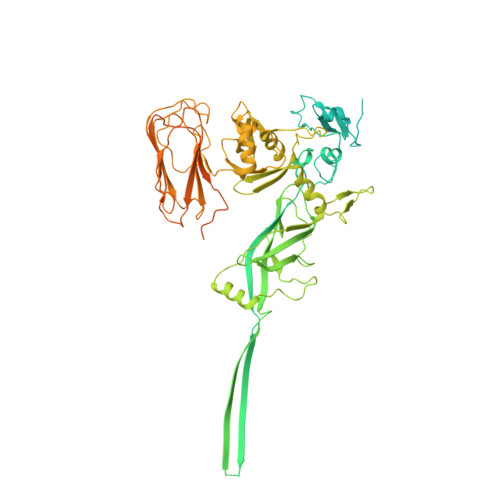
Structural insights into the transition of Clostridioides difficile binary toxin from prepore to pore.
Anderson, D.M., Sheedlo, M.J., Jensen, J.L., Lacy, D.B.(2020) Nat Microbiol 5: 102-107
- PubMed: 31712627 Search on PubMedSearch on PubMed Central
- DOI: https://doi.org/10.1038/s41564-019-0601-8
- Primary Citation Related Structures:
6O2M, 6O2N, 6O2O, 6OKR, 6OKS, 6OKT, 6OKU - PubMed Abstract:
Clostridioides (formerly Clostridium) difficile is a Gram-positive, spore-forming anaerobe and a leading cause of hospital-acquired infection and gastroenteritis-associated death in US hospitals 1 . The disease state is usually preceded by disruption of the host microbiome in response to antibiotic treatment and is characterized by mild to severe diarrhoea. C. difficile infection is dependent on the secretion of one or more AB-type toxins: toxin A (TcdA), toxin B (TcdB) and the C. difficile transferase toxin (CDT) 2 . Whereas TcdA and TcdB are considered the primary virulence factors, recent studies suggest that CDT increases the severity of C. difficile infection in some of the most problematic clinical strains 3 . To better understand how CDT functions, we used cryo-electron microscopy to define the structure of CDTb, the cell-binding component of CDT. We obtained structures of several oligomeric forms that highlight the conformational changes that enable conversion from a prepore to a β-barrel pore. The structural analysis also reveals a glycan-binding domain and residues involved in binding the host-cell receptor, lipolysis-stimulated lipoprotein receptor. Together, these results provide a framework to understand how CDT functions at the host cell interface.
- Department of Pathology, Microbiology and Immunology, Vanderbilt University Medical Center, Nashville, TN, USA.
Organizational Affiliation: