6OIV

XFEL structure of Escherichia coli dGTPase


Experimental Data Snapshot

  • Method: X-RAY DIFFRACTION
  • Resolution: 3.06 Å
  • R-Value Free: 0.248 
  • R-Value Work: 0.225 
  • R-Value Observed: 0.226 

wwPDB Validation   3D Report Full Report


This is version 1.3 of the entry. See complete history


Literature

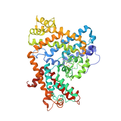
The crystal structure of dGTPase reveals the molecular basis of dGTP selectivity.

Barnes, C.O.Wu, Y.Song, J.Lin, G.Baxter, E.L.Brewster, A.S.Nagarajan, V.Holmes, A.Soltis, S.M.Sauter, N.K.Ahn, J.Cohen, A.E.Calero, G.

(2019) Proc Natl Acad Sci U S A 116: 9333-9339

  • DOI: https://doi.org/10.1073/pnas.1814999116
  • Primary Citation of Related Structures:  
    6OI7, 6OIV, 6OIW, 6OIX, 6OIY

  • PubMed Abstract: 

    Deoxynucleotide triphosphohydrolases (dNTPases) play a critical role in cellular survival and DNA replication through the proper maintenance of cellular dNTP pools. While the vast majority of these enzymes display broad activity toward canonical dNTPs, such as the dNTPase SAMHD1 that blocks reverse transcription of retroviruses in macrophages by maintaining dNTP pools at low levels, Escherichia coli ( Ec ) - dGTPase is the only known enzyme that specifically hydrolyzes dGTP. However, the mechanism behind dGTP selectivity is unclear. Here we present the free-, ligand (dGTP)- and inhibitor (GTP)-bound structures of hexameric Ec- dGTPase, including an X-ray free-electron laser structure of the free Ec -dGTPase enzyme to 3.2 Å. To obtain this structure, we developed a method that applied UV-fluorescence microscopy, video analysis, and highly automated goniometer-based instrumentation to map and rapidly position individual crystals randomly located on fixed target holders, resulting in the highest indexing rates observed for a serial femtosecond crystallography experiment. Our structures show a highly dynamic active site where conformational changes are coupled to substrate (dGTP), but not inhibitor binding, since GTP locks dGTPase in its apo- form. Moreover, despite no sequence homology, Ec -dGTPase and SAMHD1 share similar active-site and HD motif architectures; however, Ec -dGTPase residues at the end of the substrate-binding pocket mimic Watson-Crick interactions providing guanine base specificity, while a 7-Å cleft separates SAMHD1 residues from dNTP bases, abolishing nucleotide-type discrimination. Furthermore, the structures shed light on the mechanism by which long distance binding (25 Å) of single-stranded DNA in an allosteric site primes the active site by conformationally "opening" a tyrosine gate allowing enhanced substrate binding.


  • Organizational Affiliation

    Department of Structural Biology, University of Pittsburgh School of Medicine, Pittsburgh, PA 15260.


Macromolecules
Find similar proteins by:  (by identity cutoff)  |  3D Structure
Entity ID: 1
MoleculeChains Sequence LengthOrganismDetailsImage
Deoxyguanosinetriphosphate triphosphohydrolase
A, B, C, D, E
A, B, C, D, E, F
505Escherichia coli K-12Mutation(s): 0 
Gene Names: dgtb0160JW0156
EC: 3.1.5.1
UniProt
Find proteins for P15723 (Escherichia coli (strain K12))
Explore P15723 
Go to UniProtKB:  P15723
Entity Groups  
Sequence Clusters30% Identity50% Identity70% Identity90% Identity95% Identity100% Identity
UniProt GroupP15723
Sequence Annotations
Expand
  • Reference Sequence
Small Molecules
Ligands 2 Unique
IDChains Name / Formula / InChI Key2D Diagram3D Interactions
SO4
Query on SO4

Download Ideal Coordinates CCD File 
H [auth A]
J [auth B]
L [auth C]
M [auth C]
N [auth C]
H [auth A],
J [auth B],
L [auth C],
M [auth C],
N [auth C],
O [auth C],
Q [auth D],
S [auth E],
U [auth F]
SULFATE ION
O4 S
QAOWNCQODCNURD-UHFFFAOYSA-L
MN
Query on MN

Download Ideal Coordinates CCD File 
G [auth A]
I [auth B]
K [auth C]
P [auth D]
R [auth E]
G [auth A],
I [auth B],
K [auth C],
P [auth D],
R [auth E],
T [auth F]
MANGANESE (II) ION
Mn
WAEMQWOKJMHJLA-UHFFFAOYSA-N
Modified Residues  1 Unique
IDChains TypeFormula2D DiagramParent
MSE
Query on MSE
A, B, C, D, E
A, B, C, D, E, F
L-PEPTIDE LINKINGC5 H11 N O2 SeMET
Experimental Data & Validation

Experimental Data

  • Method: X-RAY DIFFRACTION
  • Resolution: 3.06 Å
  • R-Value Free: 0.248 
  • R-Value Work: 0.225 
  • R-Value Observed: 0.226 
  • Space Group: P 43 21 2
Unit Cell:
Length ( Å )Angle ( ˚ )
a = 192.589α = 90
b = 192.589β = 90
c = 291.254γ = 90
Software Package:
Software NamePurpose
BUSTERrefinement
cctbx.xfeldata reduction
cctbx.primedata scaling
PHASERphasing

Structure Validation

View Full Validation Report


Entry History & Funding Information

Deposition Data


Funding OrganizationLocationGrant Number
National Institutes of Health/National Institute of General Medical Sciences (NIH/NIGMS)United StatesR01GM112686
National Institutes of Health/National Institute of General Medical Sciences (NIH/NIGMS)United StatesR01GM116642

Revision History  (Full details and data files)

  • Version 1.0: 2019-06-05
    Type: Initial release
  • Version 1.1: 2020-01-01
    Changes: Author supporting evidence
  • Version 1.2: 2023-10-11
    Changes: Data collection, Database references, Derived calculations, Refinement description
  • Version 1.3: 2023-11-15
    Changes: Data collection