6OEZ

Crystal structure of Trypanothione Reductase from Trypanosoma brucei in complex with inhibitor (+)-N-(Cyclobutylmethyl)-3-{5-[1-(pyrrolidin-1-yl)cyclohexyl]-2-(1-{[(2S)-pyrro-lidin-2-yl]methyl}-1H-indol-5-yl)-1,3-thiazol-4-yl}prop-2-yn-1-amine


Experimental Data Snapshot

  • Method: X-RAY DIFFRACTION
  • Resolution: 2.50 Å
  • R-Value Free: 0.235 
  • R-Value Work: 0.217 
  • R-Value Observed: 0.218 

wwPDB Validation   3D Report Full Report

Currently 6OEZ does not have a validation slider image.


This is version 1.4 of the entry. See complete history


Literature

Targeting a Large Active Site: Structure-Based Design of Nanomolar Inhibitors of Trypanosoma brucei Trypanothione Reductase.

De Gasparo, R.Halgas, O.Harangozo, D.Kaiser, M.Pai, E.F.Krauth-Siegel, R.L.Diederich, F.

(2019) Chemistry 25: 11416-11421

  • DOI: https://doi.org/10.1002/chem.201901664
  • Primary Citation of Related Structures:  
    6OEX, 6OEY, 6OEZ

  • PubMed Abstract: 

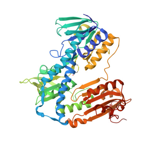
    Trypanothione reductase (TR) plays a key role in the unique redox metabolism of trypanosomatids, the causative agents of human African trypanosomiasis (HAT), Chagas' disease, and leishmaniases. Introduction of a new, lean propargylic vector to a known class of TR inhibitors resulted in the strongest reported competitive inhibitor of Trypanosoma (T.) brucei TR, with an inhibition constant K i of 73 nm, which is fully selective against human glutathione reductase (hGR). The best ligands exhibited in vitro IC 50 values (half-maximal inhibitory concentration) against the HAT pathogen, T. brucei rhodesiense, in the mid-nanomolar range, reaching down to 50 nm. X-Ray co-crystal structures confirmed the binding mode of the ligands and revealed the presence of a HEPES buffer molecule in the large active site. Extension of the propargylic vector, guided by structure-based design, to replace the HEPES buffer molecule should give inhibitors with low nanomolar K i and IC 50 values for in vivo studies.


  • Organizational Affiliation

    Laboratorium für Organische Chemie, ETH Zurich, Vladimir-Prelog-Weg 3, 8093, Zurich, Switzerland.


Macromolecules
Find similar proteins by:  (by identity cutoff)  |  3D Structure
Entity ID: 1
MoleculeChains Sequence LengthOrganismDetailsImage
Trypanothione reductaseA [auth B],
B [auth A]
495Trypanosoma brucei brucei TREU927Mutation(s): 0 
Gene Names: Tb10.406.0520
EC: 1.8.1.12
UniProt
Find proteins for Q389T8 (Trypanosoma brucei brucei (strain 927/4 GUTat10.1))
Explore Q389T8 
Go to UniProtKB:  Q389T8
Entity Groups  
Sequence Clusters30% Identity50% Identity70% Identity90% Identity95% Identity100% Identity
UniProt GroupQ389T8
Sequence Annotations
Expand
  • Reference Sequence
Small Molecules
Binding Affinity Annotations 
IDSourceBinding Affinity
M9J Binding MOAD:  6OEZ Ki: 73 (nM) from 1 assay(s)
Experimental Data & Validation

Experimental Data

  • Method: X-RAY DIFFRACTION
  • Resolution: 2.50 Å
  • R-Value Free: 0.235 
  • R-Value Work: 0.217 
  • R-Value Observed: 0.218 
  • Space Group: P 43 21 2
Unit Cell:
Length ( Å )Angle ( ˚ )
a = 117.526α = 90
b = 117.526β = 90
c = 224.522γ = 90
Software Package:
Software NamePurpose
PHENIXrefinement
CrysalisProdata reduction
Aimlessdata scaling
PHASERphasing

Structure Validation

View Full Validation Report

Currently 6OEZ does not have a validation slider image.


Entry History & Funding Information

Deposition Data


Funding OrganizationLocationGrant Number
Natural Sciences and Engineering Research Council (NSERC, Canada)CanadaRGPIN-2015-04877

Revision History  (Full details and data files)

  • Version 1.0: 2019-07-31
    Type: Initial release
  • Version 1.1: 2019-08-28
    Changes: Data collection, Database references
  • Version 1.2: 2019-09-11
    Changes: Data collection, Database references
  • Version 1.3: 2020-01-08
    Changes: Author supporting evidence
  • Version 1.4: 2023-10-11
    Changes: Data collection, Database references, Refinement description