6OCK

Crystal Structure of Leporine Serum Albumin in Complex with Ketoprofen


Experimental Data Snapshot

  • Method: X-RAY DIFFRACTION
  • Resolution: 1.90 Å
  • R-Value Free: 0.253 
  • R-Value Work: 0.190 
  • R-Value Observed: 0.192 

wwPDB Validation   3D Report Full Report

Currently 6OCK does not have a validation slider image.


This is version 1.2 of the entry. See complete history

Re-refinement Note

A newer entry is available that reflects an alternative modeling of the original data: 8BSG


Literature

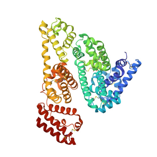
Structural investigations of stereoselective profen binding by equine and leporine serum albumins.

Zielinski, K.Sekula, B.Bujacz, A.Szymczak, I.

(2020) Chirality 32: 334-344

  • DOI: https://doi.org/10.1002/chir.23162
  • Primary Citation of Related Structures:  
    6OCI, 6OCJ, 6OCK, 6OCL

  • PubMed Abstract: 

    Serum albumin, the most abundant transport protein of mammalian blood, interacts with various nonsteroidal anti-inflammatory drugs (NSAIDs) affecting their disposition, metabolism, and excretion. A big group of chiral NSAIDs transported by albumin, profens, is created by derivatives of 2-arylpropionic acid. The chiral center in the structures of profens is adjacent to the carboxylate moiety and often determines different pharmacological properties of profen enantiomers. This study describes crystal structures of two albumins, isolated from equine and leporine serum, in complexes with three profens: ibuprofen, ketoprofen, and suprofen. Based on three-dimensional structures, the stereoselectivity of albumin is discussed and referred to the previously published albumin complexes with drugs. Drug Site 2 (DS2) of albumin, the bulky hydrophobic pocket of subdomain IIIA with a patch of polar residues, preferentially binds (S)-enantiomers of all investigated profens. Almost identical binding mode of all these drugs clearly indicates the stereoselectivity of DS2 towards (S)-profens in different albumin species. Also, the affinity studies show that DS2 is the major site that presents high affinity towards investigated drugs. Additionally, crystallographic data reveal the secondary binding sites of ketoprofen in leporine serum albumin and ibuprofen in equine serum albumin, both overlapping with previously identified naproxen binding sites: the cleft formed between subdomains IIIA and IIIB close to the fatty acid binding site 5 and the niche created between subdomains IIA and IIIA, called fatty acid site 6.


  • Organizational Affiliation

    Institute of Molecular and Industrial Biotechnology, Lodz University of Technology, Lodz, Poland.


Macromolecules
Find similar proteins by:  (by identity cutoff)  |  3D Structure
Entity ID: 1
MoleculeChains Sequence LengthOrganismDetailsImage
Serum albumin584Oryctolagus cuniculusMutation(s): 0 
UniProt
Find proteins for P49065 (Oryctolagus cuniculus)
Explore P49065 
Go to UniProtKB:  P49065
Entity Groups  
Sequence Clusters30% Identity50% Identity70% Identity90% Identity95% Identity100% Identity
UniProt GroupP49065
Sequence Annotations
Expand
  • Reference Sequence
Small Molecules
Ligands 5 Unique
IDChains Name / Formula / InChI Key2D Diagram3D Interactions
POG
Query on POG

Download Ideal Coordinates CCD File 
J [auth A](20S)-2,5,8,11,14,17-HEXAMETHYL-3,6,9,12,15,18-HEXAOXAHENICOSANE-1,20-DIOL
C21 H44 O8
OWRNLGZKEZSHGO-QAWAPDBNSA-N
9KL
Query on 9KL

Download Ideal Coordinates CCD File 
B [auth A],
C [auth A]
(2S)-2-[3-(benzenecarbonyl)phenyl]propanoic acid
C16 H14 O3
DKYWVDODHFEZIM-NSHDSACASA-N
2J3
Query on 2J3

Download Ideal Coordinates CCD File 
K [auth A],
L [auth A]
(2R)-2-{[(2R)-2-{[(2R)-2-hydroxypropyl]oxy}propyl]oxy}propan-1-ol
C9 H20 O4
LCZVSXRMYJUNFX-IWSPIJDZSA-N
PGE
Query on PGE

Download Ideal Coordinates CCD File 
I [auth A]TRIETHYLENE GLYCOL
C6 H14 O4
ZIBGPFATKBEMQZ-UHFFFAOYSA-N
ACT
Query on ACT

Download Ideal Coordinates CCD File 
D [auth A],
E [auth A],
F [auth A],
G [auth A],
H [auth A]
ACETATE ION
C2 H3 O2
QTBSBXVTEAMEQO-UHFFFAOYSA-M
Binding Affinity Annotations 
IDSourceBinding Affinity
9KL Binding MOAD:  6OCK Ka: 2.04e+4 (M^-1) from 1 assay(s)
Experimental Data & Validation

Experimental Data

  • Method: X-RAY DIFFRACTION
  • Resolution: 1.90 Å
  • R-Value Free: 0.253 
  • R-Value Work: 0.190 
  • R-Value Observed: 0.192 
  • Space Group: P 21 21 21
Unit Cell:
Length ( Å )Angle ( ˚ )
a = 73.69α = 90
b = 79.48β = 90
c = 103.84γ = 90
Software Package:
Software NamePurpose
REFMACrefinement
XDSdata reduction
XSCALEdata scaling
PHASERphasing

Structure Validation

View Full Validation Report

Currently 6OCK does not have a validation slider image.


Entry History 

Deposition Data

Revision History  (Full details and data files)

  • Version 1.0: 2020-01-15
    Type: Initial release
  • Version 1.1: 2020-02-26
    Changes: Database references
  • Version 1.2: 2023-10-11
    Changes: Data collection, Database references, Refinement description, Structure summary