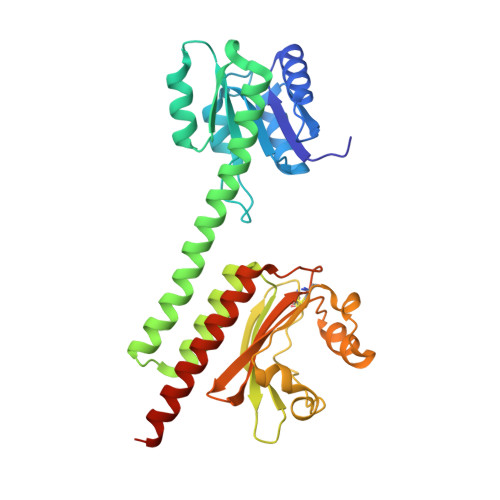
Structural basis of molecular logic OR in a dual-sensor histidine kinase.
Shin, H., Ren, Z., Zeng, X., Bandara, S., Yang, X.(2019) Proc Natl Acad Sci U S A 116: 19973-19982
- PubMed: 31527275 Search on PubMedSearch on PubMed Central
- DOI: https://doi.org/10.1073/pnas.1910855116
- Primary Citation Related Structures:
6OAP, 6OAQ, 6OB8 - PubMed Abstract:
Signal detection and integration by sensory proteins constitute the critical molecular events as living organisms respond to changes in a complex environment. Many sensory proteins adopt a modular architecture that integrates the perception of distinct chemical or physical signals and the generation of a biological response in the same protein molecule. Currently, how signal perception and integration are achieved in such a modular, often dimeric, framework remains elusive. Here, we report a dynamic crystallography study on the tandem sensor domains of a dual-sensor histidine kinase PPHK (phosphorylation-responsive photosensitive histidine kinase) that operates a molecular logic OR, by which the output kinase activity is modulated by a phosphorylation signal and a light signal. A joint analysis of ∼170 crystallographic datasets probing different signaling states shows remarkable dimer asymmetry as PPHK responds to the input signals and transitions from one state to the other. Supported by mutational data and structural analysis, these direct observations reveal the working mechanics of the molecular logic OR in PPHK, where the light-induced bending of a long signaling helix at the dimer interface is counteracted by the ligand-induced structural changes from a different sensor domain. We propose that the logic OR of PPHK, together with an upstream photoreceptor, implements a "long-pass" red light response distinct from those accomplished by classical phytochromes.
- Department of Chemistry, The University of Illinois at Chicago, Chicago, IL 60607.
Organizational Affiliation: