


Mycobacterium tuberculosis L-alanine dehydrogenase x-ray structure in complex with N6-isobutyl adenosine
Kim, H.-B., Hung, L.-W., Terwilliger, T.C., Kim, C.-Y.To be published.
Experimental Data Snapshot
Entity ID: 1 | |||||
|---|---|---|---|---|---|
| Molecule | Chains | Sequence Length | Organism | Details | Image |
| Alanine dehydrogenase | 371 | Mycobacterium tuberculosis H37Rv | Mutation(s): 0 Gene Names: ald, Rv2780, MTV002.45 EC: 1.4.1.1 | 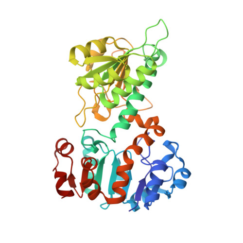 | |
UniProt | |||||
Entity Groups | |||||
| Sequence Clusters | 30% Identity50% Identity70% Identity90% Identity95% Identity100% Identity | ||||
| UniProt Group | P9WQB1 | ||||
Sequence AnnotationsExpand | |||||
Reference Sequence | |||||
| Ligands 1 Unique | |||||
|---|---|---|---|---|---|
| ID | Chains | Name / Formula / InChI Key | 2D Diagram | 3D Interactions | |
| NKV Download:Ideal Coordinates CCD File | B [auth A] | N-(2-methylpropanoyl)adenosine C14 H19 N5 O5 RSLAEWMJDPSZPS-AKAIJSEGSA-N |  | ||
| Length ( Å ) | Angle ( ˚ ) |
|---|---|
| a = 88.423 | α = 90 |
| b = 88.423 | β = 90 |
| c = 289.991 | γ = 120 |
| Software Name | Purpose |
|---|---|
| PHENIX | refinement |
| HKL-2000 | data reduction |
| HKL-2000 | data scaling |
| PHENIX | phasing |


