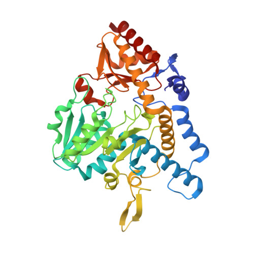
6O10

E. coli cysteine desulfurase SufS


Experimental Data Snapshot

  • Method: X-RAY DIFFRACTION
  • Resolution: 2.00 Å
  • R-Value Free: 0.204 
  • R-Value Work: 0.182 
  • R-Value Observed: 0.183 

Starting Model: experimental
View more details

wwPDB Validation   3D Report Full Report


Ligand Structure Quality Assessment 


This is version 1.4 of the entry. See complete history


Literature

Direct observation of intermediates in the SufS cysteine desulfurase reaction reveals functional roles of conserved active-site residues.

Blahut, M.Wise, C.E.Bruno, M.R.Dong, G.Makris, T.M.Frantom, P.A.Dunkle, J.A.Outten, F.W.

(2019) J Biol Chem 294: 12444-12458

  • DOI: https://doi.org/10.1074/jbc.RA119.009471
  • Primary Citation of Related Structures:  
    6O10, 6O11, 6O12, 6O13

  • PubMed Abstract: 

    Iron-sulfur (Fe-S) clusters are necessary for the proper functioning of numerous metalloproteins. Fe-S cluster (Isc) and sulfur utilization factor (Suf) pathways are the key biosynthetic routes responsible for generating these Fe-S cluster prosthetic groups in Escherichia coli Although Isc dominates under normal conditions, Suf takes over during periods of iron depletion and oxidative stress. Sulfur acquisition via these systems relies on the ability to remove sulfur from free cysteine using a cysteine desulfurase mechanism. In the Suf pathway, the dimeric SufS protein uses the cofactor pyridoxal 5'-phosphate (PLP) to abstract sulfur from free cysteine, resulting in the production of alanine and persulfide. Despite much progress, the stepwise mechanism by which this PLP-dependent enzyme operates remains unclear. Here, using rapid-mixing kinetics in conjunction with X-ray crystallography, we analyzed the pre-steady-state kinetics of this process while assigning early intermediates of the mechanism. We employed H123A and C364A SufS variants to trap Cys-aldimine and Cys-ketimine intermediates of the cysteine desulfurase reaction, enabling direct observations of these intermediates and associated conformational changes of the SufS active site. Of note, we propose that Cys-364 is essential for positioning the Cys-aldimine for Cα deprotonation, His-123 acts to protonate the Ala-enamine intermediate, and Arg-56 facilitates catalysis by hydrogen bonding with the sulfhydryl of Cys-aldimine. Our results, along with previous SufS structural findings, suggest a detailed model of the SufS-catalyzed reaction from Cys binding to C-S bond cleavage and indicate that Arg-56, His-123, and Cys-364 are critical SufS residues in this C-S bond cleavage pathway.


  • Organizational Affiliation

    Department of Chemistry and Biochemistry, University of South Carolina, Columbia, South Carolina 29208.


Macromolecules
Find similar proteins by:  (by identity cutoff)  |  3D Structure
Entity ID: 1
MoleculeChains Sequence LengthOrganismDetailsImage
Cysteine desulfurase406Escherichia coli K-12Mutation(s): 0 
Gene Names: sufScsdBynhBb1680JW1670
EC: 2.8.1.7 (PDB Primary Data), 4.4.1.16 (PDB Primary Data)
UniProt
Find proteins for P77444 (Escherichia coli (strain K12))
Explore P77444 
Go to UniProtKB:  P77444
Entity Groups  
Sequence Clusters30% Identity50% Identity70% Identity90% Identity95% Identity100% Identity
UniProt GroupP77444
Sequence Annotations
Expand
  • Reference Sequence
Small Molecules
Ligands 2 Unique
IDChains Name / Formula / InChI Key2D Diagram3D Interactions
PLP
Query on PLP

Download Ideal Coordinates CCD File 
B [auth A]PYRIDOXAL-5'-PHOSPHATE
C8 H10 N O6 P
NGVDGCNFYWLIFO-UHFFFAOYSA-N
CL
Query on CL

Download Ideal Coordinates CCD File 
C [auth A]CHLORIDE ION
Cl
VEXZGXHMUGYJMC-UHFFFAOYSA-M
Experimental Data & Validation

Experimental Data

  • Method: X-RAY DIFFRACTION
  • Resolution: 2.00 Å
  • R-Value Free: 0.204 
  • R-Value Work: 0.182 
  • R-Value Observed: 0.183 
  • Space Group: P 43 21 2
Unit Cell:
Length ( Å )Angle ( ˚ )
a = 126.4α = 90
b = 126.4β = 90
c = 133.68γ = 90
Software Package:
Software NamePurpose
XSCALEdata scaling
PHENIXrefinement
PDB_EXTRACTdata extraction
XDSdata reduction
PHENIXphasing

Structure Validation

View Full Validation Report


Ligand Structure Quality Assessment 


Entry History & Funding Information

Deposition Data


Funding OrganizationLocationGrant Number
National Institutes of Health/National Institute of General Medical Sciences (NIH/NIGMS)United StatesGM112919

Revision History  (Full details and data files)

  • Version 1.0: 2019-06-26
    Type: Initial release
  • Version 1.1: 2019-07-10
    Changes: Data collection, Database references
  • Version 1.2: 2019-08-28
    Changes: Data collection, Database references
  • Version 1.3: 2020-01-01
    Changes: Author supporting evidence
  • Version 1.4: 2023-10-11
    Changes: Advisory, Data collection, Database references, Refinement description