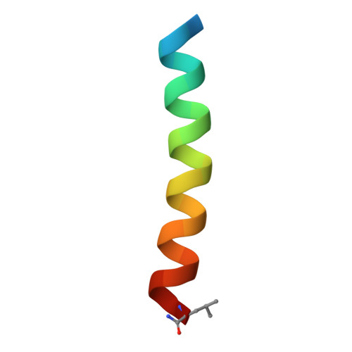
6NV1

Structure of drug-resistant V27A mutant of the influenza M2 proton channel bound to spiroadamantyl amine inhibitor


Experimental Data Snapshot

  • Method: X-RAY DIFFRACTION
  • Resolution: 2.50 Å
  • R-Value Free: 0.258 
  • R-Value Work: 0.220 
  • R-Value Observed: 0.224 

Starting Model: experimental
View more details

wwPDB Validation   3D Report Full Report


Ligand Structure Quality Assessment 


This is version 1.3 of the entry. See complete history


Literature

X-ray Crystal Structures of the Influenza M2 Proton Channel Drug-Resistant V27A Mutant Bound to a Spiro-Adamantyl Amine Inhibitor Reveal the Mechanism of Adamantane Resistance.

Thomaston, J.L.Konstantinidi, A.Liu, L.Lambrinidis, G.Tan, J.Caffrey, M.Wang, J.Degrado, W.F.Kolocouris, A.

(2020) Biochemistry 59: 627-634

  • DOI: https://doi.org/10.1021/acs.biochem.9b00971
  • Primary Citation of Related Structures:  
    6NV1, 6OUG

  • PubMed Abstract: 

    The V27A mutation confers adamantane resistance on the influenza A matrix 2 (M2) proton channel and is becoming more prevalent in circulating populations of influenza A virus. We have used X-ray crystallography to determine structures of a spiro-adamantyl amine inhibitor bound to M2(22-46) V27A and also to M2(21-61) V27A in the Inward closed conformation. The spiro-adamantyl amine binding site is nearly identical for the two crystal structures. Compared to the M2 "wild type" (WT) with valine at position 27, we observe that the channel pore is wider at its N-terminus as a result of the V27A mutation and that this removes V27 side chain hydrophobic interactions that are important for binding of amantadine and rimantadine. The spiro-adamantyl amine inhibitor blocks proton conductance in the WT and V27A mutant channels by shifting its binding site in the pore depending on which residue is present at position 27. Additionally, in the structure of the M2(21-61) V27A construct, the C-terminus of the channel is tightly packed relative to that of the M2(22-46) construct. We observe that residues Asp44, Arg45, and Phe48 face the center of the channel pore and would be well-positioned to interact with protons exiting the M2 channel after passing through the His37 gate. A 300 ns molecular dynamics simulation of the M2(22-46) V27A-spiro-adamantyl amine complex predicts with accuracy the position of the ligands and waters inside the pore in the X-ray crystal structure of the M2(22-46) V27A complex.


  • Organizational Affiliation

    Department of Pharmaceutical Chemistry , University of California San Francisco , San Francisco , California 94158 , United States.


Macromolecules
Find similar proteins by:  (by identity cutoff)  |  3D Structure
Entity ID: 1
MoleculeChains Sequence LengthOrganismDetailsImage
Matrix protein 2
A, B, C, D, E
A, B, C, D, E, F, G, H
27Influenza A virus (A/Indonesia/CDC1031RE2/2007(H5N1))Mutation(s): 2 
Membrane Entity: Yes 
UniProt
Find proteins for A4D7H3 (Influenza A virus)
Explore A4D7H3 
Go to UniProtKB:  A4D7H3
Entity Groups  
Sequence Clusters30% Identity50% Identity70% Identity90% Identity95% Identity100% Identity
UniProt GroupA4D7H3
Sequence Annotations
Expand
  • Reference Sequence
Small Molecules
Ligands 3 Unique
IDChains Name / Formula / InChI Key2D Diagram3D Interactions
OLC
Query on OLC

Download Ideal Coordinates CCD File 
I [auth A]
K [auth B]
M [auth D]
N [auth E]
O [auth E]
I [auth A],
K [auth B],
M [auth D],
N [auth E],
O [auth E],
P [auth F],
R [auth G],
S [auth H]
(2R)-2,3-dihydroxypropyl (9Z)-octadec-9-enoate
C21 H40 O4
RZRNAYUHWVFMIP-GDCKJWNLSA-N
E01
Query on E01

Download Ideal Coordinates CCD File 
J [auth A],
Q [auth F]
(1r,1'S,3'S,5'S,7'S)-spiro[cyclohexane-1,2'-tricyclo[3.3.1.1~3,7~]decan]-4-amine
C15 H25 N
VNTMIVFMVGTSHV-RLUXGHLOSA-N
CL
Query on CL

Download Ideal Coordinates CCD File 
L [auth B],
T [auth H]
CHLORIDE ION
Cl
VEXZGXHMUGYJMC-UHFFFAOYSA-M
Experimental Data & Validation

Experimental Data

  • Method: X-RAY DIFFRACTION
  • Resolution: 2.50 Å
  • R-Value Free: 0.258 
  • R-Value Work: 0.220 
  • R-Value Observed: 0.224 
  • Space Group: P 21 21 21
Unit Cell:
Length ( Å )Angle ( ˚ )
a = 49.806α = 90
b = 49.95β = 90
c = 75.029γ = 90
Software Package:
Software NamePurpose
PHENIXrefinement
XDSdata reduction
XDSdata scaling
PHASERphasing

Structure Validation

View Full Validation Report


Ligand Structure Quality Assessment 


Entry History & Funding Information

Deposition Data


Funding OrganizationLocationGrant Number
National Institutes of Health/National Human Genome Research Institute (NIH/NHGRI)United StatesR01-GM056423

Revision History  (Full details and data files)

  • Version 1.0: 2020-01-15
    Type: Initial release
  • Version 1.1: 2020-02-19
    Changes: Database references
  • Version 1.2: 2023-10-11
    Changes: Data collection, Database references, Refinement description
  • Version 1.3: 2024-10-23
    Changes: Structure summary