6NT6

Cryo-EM structure of full-length chicken STING in the apo state


Experimental Data Snapshot

  • Method: ELECTRON MICROSCOPY
  • Resolution: 4.00 Å
  • Aggregation State: PARTICLE 
  • Reconstruction Method: SINGLE PARTICLE 

wwPDB Validation   3D Report Full Report


This is version 1.5 of the entry. See complete history


Literature

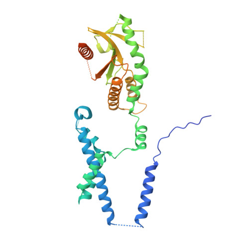
Cryo-EM structures of STING reveal its mechanism of activation by cyclic GMP-AMP.

Shang, G.Zhang, C.Chen, Z.J.Bai, X.C.Zhang, X.

(2019) Nature 567: 389-393

  • DOI: https://doi.org/10.1038/s41586-019-0998-5
  • Primary Citation of Related Structures:  
    6NT5, 6NT6, 6NT7, 6NT8

  • PubMed Abstract: 

    Infections by pathogens that contain DNA trigger the production of type-I interferons and inflammatory cytokines through cyclic GMP-AMP synthase, which produces 2'3'-cyclic GMP-AMP (cGAMP) that binds to and activates stimulator of interferon genes (STING; also known as TMEM173, MITA, ERIS and MPYS) 1-8 . STING is an endoplasmic-reticulum membrane protein that contains four transmembrane helices followed by a cytoplasmic ligand-binding and signalling domain 9-13 . The cytoplasmic domain of STING forms a dimer, which undergoes a conformational change upon binding to cGAMP 9,14 . However, it remains unclear how this conformational change leads to STING activation. Here we present cryo-electron microscopy structures of full-length STING from human and chicken in the inactive dimeric state (about 80 kDa in size), as well as cGAMP-bound chicken STING in both the dimeric and tetrameric states. The structures show that the transmembrane and cytoplasmic regions interact to form an integrated, domain-swapped dimeric assembly. Closure of the ligand-binding domain, induced by cGAMP, leads to a 180° rotation of the ligand-binding domain relative to the transmembrane domain. This rotation is coupled to a conformational change in a loop on the side of the ligand-binding-domain dimer, which leads to the formation of the STING tetramer and higher-order oligomers through side-by-side packing. This model of STING oligomerization and activation is supported by our structure-based mutational analyses.


  • Organizational Affiliation

    Department of Pharmacology, University of Texas Southwestern Medical Center, Dallas, TX, USA.


Macromolecules
Find similar proteins by:  (by identity cutoff)  |  3D Structure
Entity ID: 1
MoleculeChains Sequence LengthOrganismDetailsImage
Stimulator of interferon genes protein
A, B
392Gallus gallusMutation(s): 0 
Gene Names: TMEM173
Membrane Entity: Yes 
UniProt
Find proteins for E1C7U0 (Gallus gallus)
Explore E1C7U0 
Go to UniProtKB:  E1C7U0
Entity Groups  
Sequence Clusters30% Identity50% Identity70% Identity90% Identity95% Identity100% Identity
UniProt GroupE1C7U0
Sequence Annotations
Expand
  • Reference Sequence
Experimental Data & Validation

Experimental Data

  • Method: ELECTRON MICROSCOPY
  • Resolution: 4.00 Å
  • Aggregation State: PARTICLE 
  • Reconstruction Method: SINGLE PARTICLE 
EM Software:
TaskSoftware PackageVersion
MODEL REFINEMENTPHENIX
RECONSTRUCTIONRELION

Structure Validation

View Full Validation Report


Entry History & Funding Information

Deposition Data


Funding OrganizationLocationGrant Number
National Institutes of Health/National Institute of General Medical Sciences (NIH/NIGMS)United StatesGM088197
National Institutes of Health/National Institute of General Medical Sciences (NIH/NIGMS)United StatesR35GM130289
Howard Hughes Medical Institute (HHMI)United States--
Welch FoundationUnited StatesI-1389
Welch FoundationUnited StatesI-1702
Welch FoundationUnited StatesI-1944

Revision History  (Full details and data files)

  • Version 1.0: 2019-03-06
    Type: Initial release
  • Version 1.1: 2019-03-20
    Changes: Data collection, Database references
  • Version 1.2: 2019-04-03
    Changes: Data collection, Database references
  • Version 1.3: 2019-11-20
    Changes: Author supporting evidence
  • Version 1.4: 2019-12-18
    Changes: Other
  • Version 1.5: 2024-03-20
    Changes: Data collection, Database references