6NPT

TRK-A IN COMPLEX WITH LIGAND 1


Experimental Data Snapshot

  • Method: X-RAY DIFFRACTION
  • Resolution: 2.19 Å
  • R-Value Free: 0.276 
  • R-Value Work: 0.223 
  • R-Value Observed: 0.227 

wwPDB Validation   3D Report Full Report


Ligand Structure Quality Assessment 


This is version 1.1 of the entry. See complete history


Literature

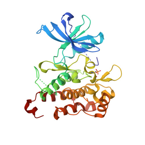
Deciphering the Allosteric Binding Mechanism of the Human Tropomyosin Receptor Kinase A ( hTrkA) Inhibitors.

Subramanian, G.Johnson, P.D.Zachary, T.Roush, N.Zhu, Y.Bowen, S.J.Janssen, A.Duclos, B.A.Williams, T.Javens, C.Shalaly, N.D.Molina, D.M.Wittwer, A.J.Hirsch, J.L.

(2019) ACS Chem Biol 14: 1205-1216

  • DOI: https://doi.org/10.1021/acschembio.9b00126
  • Primary Citation of Related Structures:  
    6NPT, 6NSP, 6NSS

  • PubMed Abstract: 

    Access to cryptic binding pockets or allosteric sites on a kinase that present themselves when the enzyme is in a specific conformational state offers a paradigm shift in designing the next generation small molecule kinase inhibitors. The current work showcases an extensive and exhaustive array of in vitro biochemical and biophysical tools and techniques deployed along with structural biology efforts of inhibitor-bound kinase complexes to characterize and confirm the cryptic allosteric binding pocket and docking mode of the small molecule actives identified for hTrkA. Specifically, assays were designed and implemented to lock the kinase in a predominantly active or inactive conformation and the effect of the kinase inhibitor probed to understand the hTrkA binding and hTrkB selectivity. The current outcome suggests that inhibitors with a fast association rate take advantage of the inactive protein conformation and lock the kinase state by also exhibiting a slow off-rate. This in turn shifts the inactive/active state protein conformational equilibrium cycle, affecting the subsequent downstream signaling.


  • Organizational Affiliation

    Veterinary Medicine Research & Development (VMRD) , Zoetis, 333 Portage Street , Kalamazoo , Michigan 49007 , United States.


Macromolecules
Find similar proteins by:  (by identity cutoff)  |  3D Structure
Entity ID: 1
MoleculeChains Sequence LengthOrganismDetailsImage
High affinity nerve growth factor receptor305Homo sapiensMutation(s): 0 
Gene Names: NTRK1MTCTRKTRKA
EC: 2.7.10.1
UniProt & NIH Common Fund Data Resources
Find proteins for P04629 (Homo sapiens)
Explore P04629 
Go to UniProtKB:  P04629
PHAROS:  P04629
GTEx:  ENSG00000198400 
Entity Groups  
Sequence Clusters30% Identity50% Identity70% Identity90% Identity95% Identity100% Identity
UniProt GroupP04629
Sequence Annotations
Expand
  • Reference Sequence
Small Molecules
Ligands 1 Unique
IDChains Name / Formula / InChI Key2D Diagram3D Interactions
KWY
Query on KWY

Download Ideal Coordinates CCD File 
B [auth A]4-tert-butyl-N-(1,3-diphenyl-1H-pyrazol-5-yl)benzamide
C26 H25 N3 O
GTSXKUJFTQTICP-UHFFFAOYSA-N
Modified Residues  1 Unique
IDChains TypeFormula2D DiagramParent
SEP
Query on SEP
A
L-PEPTIDE LINKINGC3 H8 N O6 PSER
Binding Affinity Annotations 
IDSourceBinding Affinity
KWY Binding MOAD:  6NPT IC50: 0.07 (nM) from 1 assay(s)
Experimental Data & Validation

Experimental Data

  • Method: X-RAY DIFFRACTION
  • Resolution: 2.19 Å
  • R-Value Free: 0.276 
  • R-Value Work: 0.223 
  • R-Value Observed: 0.227 
  • Space Group: P 31 1 2
Unit Cell:
Length ( Å )Angle ( ˚ )
a = 52.084α = 90
b = 52.084β = 90
c = 225.611γ = 120
Software Package:
Software NamePurpose
XDSdata reduction
XSCALEdata scaling
REFMACrefinement
Cootmodel building

Structure Validation

View Full Validation Report


Ligand Structure Quality Assessment 


Entry History 

Deposition Data

Revision History  (Full details and data files)

  • Version 1.0: 2019-05-22
    Type: Initial release
  • Version 1.1: 2019-07-10
    Changes: Data collection, Database references