6NPI | pdb_00006npi

Crystal structure of Epstein-Barr Virus Nuclear Antigen-1, EBNA1, bound to fragments


Experimental Data Snapshot

  • Method: X-RAY DIFFRACTION
  • Resolution: 1.50 Å
  • R-Value Free: 
    0.163 (Depositor), 0.162 (DCC) 
  • R-Value Work: 
    0.141 (Depositor), 0.142 (DCC) 
  • R-Value Observed: 
    0.142 (Depositor) 

Starting Model: experimental
View more details

wwPDB Validation 3D Report Full Report

Validation slider image for 6NPI

Ligand Structure Quality Assessment 


This is version 1.1 of the entry. See complete history

Literature

Structure-based design of small-molecule inhibitors of EBNA1 DNA binding blocks Epstein-Barr virus latent infection and tumor growth.

Messick, T.E.Smith, G.R.Soldan, S.S.McDonnell, M.E.Deakyne, J.S.Malecka, K.A.Tolvinski, L.van den Heuvel, A.P.J.Gu, B.W.Cassel, J.A.Tran, D.H.Wassermann, B.R.Zhang, Y.Velvadapu, V.Zartler, E.R.Busson, P.Reitz, A.B.Lieberman, P.M.

(2019) Sci Transl Med 11

  • DOI: https://doi.org/10.1126/scitranslmed.aau5612
  • Primary Citation Related Structures: 
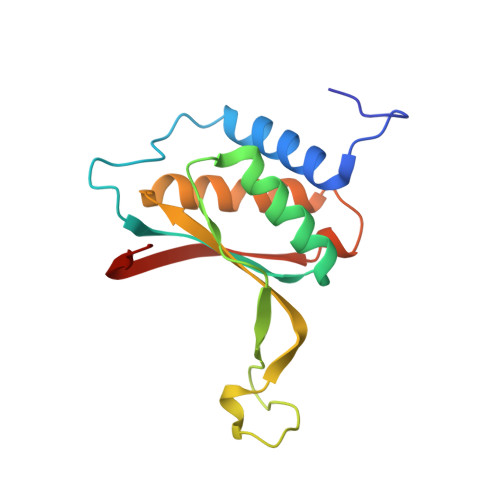
    6NPI, 6NPM, 6NPP

  • PubMed Abstract: 

    Epstein-Barr virus (EBV) is a DNA tumor virus responsible for 1 to 2% of human cancers including subtypes of Burkitt's lymphoma, Hodgkin's lymphoma, gastric carcinoma, and nasopharyngeal carcinoma (NPC). Persistent latent infection drives EBV-associated tumorigenesis. Epstein-Barr nuclear antigen 1 (EBNA1) is the only viral protein consistently expressed in all EBV-associated tumors and is therefore an attractive target for therapeutic intervention. It is a multifunctional DNA binding protein critical for viral replication, genome maintenance, viral gene expression, and host cell survival. Using a fragment-based approach and x-ray crystallography, we identify a 2,3-disubstituted benzoic acid series that selectively inhibits the DNA binding activity of EBNA1. We characterize these inhibitors biochemically and in cell-based assays, including chromatin immunoprecipitation and DNA replication assays. In addition, we demonstrate the potency of EBNA1 inhibitors to suppress tumor growth in several EBV-dependent xenograft models, including patient-derived xenografts for NPC. These inhibitors selectively block EBV gene transcription and alter the cellular transforming growth factor-β (TGF-β) signaling pathway in NPC tumor xenografts. These EBNA1-specific inhibitors show favorable pharmacological properties and have the potential to be further developed for the treatment of EBV-associated malignancies.


  • Organizational Affiliation
    • The Wistar Institute, 3601 Spruce Street, Philadelphia, PA 19104, USA. lieberman@wistar.org troymessick@gmail.com.

Macromolecule Content 

  • Total Structure Weight: 31.11 kDa 
  • Atom Count: 2,623 
  • Modeled Residue Count: 274 
  • Deposited Residue Count: 282 
  • Unique protein chains: 1

Macromolecules

Find similar proteins by:|  3D Structure
Entity ID: 1
MoleculeChains  Sequence LengthOrganismDetailsImage
Epstein-Barr nuclear antigen 1
A, B
141Human herpesvirus 4 strain B95-8Mutation(s): 0 
Gene Names: EBNA1BKRF1
EC: 3.1.21
UniProt
Find proteins for P03211 (Epstein-Barr virus (strain B95-8))
Explore P03211 
Go to UniProtKB:  P03211
Entity Groups
Sequence Clusters30% Identity50% Identity70% Identity90% Identity95% Identity100% Identity
UniProt GroupP03211
Sequence Annotations
Expand
Reference Sequence

Small Molecules

Ligands 2 Unique
IDChains Name / Formula / InChI Key2D Diagram3D Interactions
KW1

Query on KW1


Download:Ideal Coordinates CCD File
D [auth B]({2-[(4-bromo-5-methyl-1,2-oxazol-3-yl)amino]-2-oxoethyl}sulfanyl)acetic acid
C8 H9 Br N2 O4 S
CMJFFJICEDWDAG-UHFFFAOYSA-N
60Q

Query on 60Q


Download:Ideal Coordinates CCD File
C [auth B]2-pyrrol-1-ylbenzoic acid
C11 H9 N O2
GNWTWXOZRSBCOZ-UHFFFAOYSA-N

Experimental Data & Validation

Experimental Data

  • Method: X-RAY DIFFRACTION
  • Resolution: 1.50 Å
  • R-Value Free:  0.163 (Depositor), 0.162 (DCC) 
  • R-Value Work:  0.141 (Depositor), 0.142 (DCC) 
  • R-Value Observed: 0.142 (Depositor) 
Space Group: P 21 21 21
Unit Cell:
Length ( Å )Angle ( ˚ )
a = 59.704α = 90
b = 67.452β = 90
c = 69.498γ = 90
Software Package:
Software NamePurpose
PHENIXrefinement
HKL-3000data reduction
HKL-3000data scaling
PHENIXphasing

Structure Validation

View Full Validation Report


Ligand Structure Quality Assessment 


Entry History 

& Funding Information

Deposition Data


Funding OrganizationLocationGrant Number
Wellcome TrustUnited KingdomWT096496

Revision History  (Full details and data files)

  • Version 1.0: 2019-03-20
    Type: Initial release
  • Version 1.1: 2023-10-11
    Changes: Data collection, Database references, Refinement description