Engineering the PP7 Virus Capsid as a Peptide Display Platform.
Zhao, L., Kopylov, M., Potter, C.S., Carragher, B., Finn, M.G.(2019) ACS Nano 13: 4443-4454
- PubMed: 30912918 Search on PubMedSearch on PubMed Central
- DOI: https://doi.org/10.1021/acsnano.8b09683
- Primary Citation Related Structures:
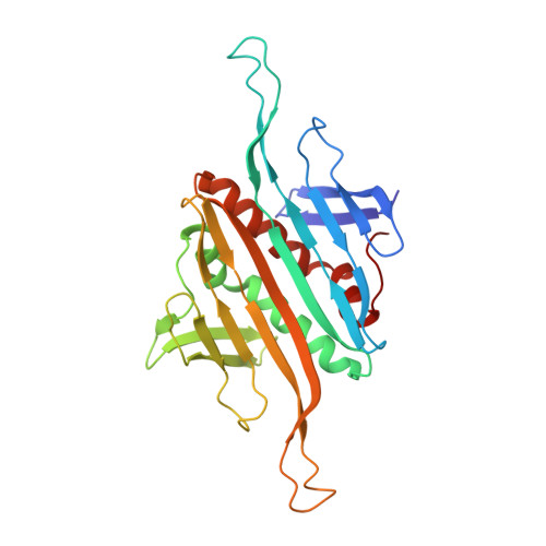
6N4V - PubMed Abstract:
As self-assembling polyvalent nanoscale structures that can tolerate substantial genetic and chemical modification, virus-like particles are useful in a variety of fields. Here we describe the genetic modification and structural characterization of the Leviviridae PP7 capsid protein as a platform for the presentation of functional polypeptides. This particle was shown to tolerate the display of sequences from 1 kDa (a cell penetrating peptide) to 14 kDa (the Fc-binding double Z-domain) on its exterior surface as C-terminal genetic fusions to the coat protein. In addition, a dimeric construct allowed the presentation of exogenous loops between capsid monomers and the simultaneous presentation of two different peptides at different positions on the icosahedral structure. The PP7 particle is thereby significantly more tolerant of these types of polypeptide additions than Qβ and MS2, the other Leviviridae-derived VLPs in common use.
- National Resource for Automated Molecular Microscopy, Simons Electron Microscopy Center, New York Structural Biology Center , 89 Convent Avenue , New York , New York 10027 , United States.
Organizational Affiliation: