Serine-47 phosphorylation of cytochromecin the mammalian brain regulates cytochromecoxidase and caspase-3 activity.
Kalpage, H.A., Vaishnav, A., Liu, J., Varughese, A., Wan, J., Turner, A.A., Ji, Q., Zurek, M.P., Kapralov, A.A., Kagan, V.E., Brunzelle, J.S., Recanati, M.A., Grossman, L.I., Sanderson, T.H., Lee, I., Salomon, A.R., Edwards, B.F.P., Huttemann, M.(2019) FASEB J 33: 13503-13514
- PubMed: 31570002 Search on PubMedSearch on PubMed Central
- DOI: https://doi.org/10.1096/fj.201901120R
- Primary Citation Related Structures:
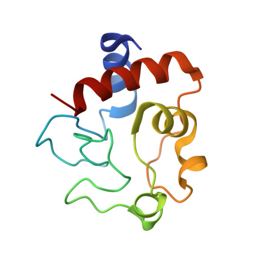
5C0Z, 6N1O - PubMed Abstract:
Cytochrome c (Cyt c ) is a multifunctional protein that operates as an electron carrier in the mitochondrial electron transport chain and plays a key role in apoptosis. We have previously shown that tissue-specific phosphorylations of Cyt c in the heart, liver, and kidney play an important role in the regulation of cellular respiration and cell death. Here, we report that Cyt c purified from mammalian brain is phosphorylated on S47 and that this phosphorylation is lost during ischemia. We have characterized the functional effects in vitro using phosphorylated Cyt c purified from pig brain tissue and a recombinant phosphomimetic mutant (S47E). We crystallized S47E phosphomimetic Cyt c at 1.55 Å and suggest that it spatially matches S47-phosphorylated Cyt c , making it a good model system. Both S47-phosphorylated and phosphomimetic Cyt c showed a lower oxygen consumption rate in reaction with isolated Cyt c oxidase, which we propose maintains intermediate mitochondrial membrane potentials under physiologic conditions, thus minimizing production of reactive oxygen species. S47-phosphorylated and phosphomimetic Cyt c showed lower caspase-3 activity. Furthermore, phosphomimetic Cyt c had decreased cardiolipin peroxidase activity and is more stable in the presence of H 2 O 2 . Our data suggest that S47 phosphorylation of Cyt c is tissue protective and promotes cell survival in the brain.-Kalpage, H. A., Vaishnav, A., Liu, J., Varughese, A., Wan, J., Turner, A. A., Ji, Q., Zurek, M. P., Kapralov, A. A., Kagan, V. E., Brunzelle, J. S., Recanati, M.-A., Grossman, L. I., Sanderson, T. H., Lee, I., Salomon, A. R., Edwards, B. F. P, Hüttemann, M. Serine-47 phosphorylation of cytochrome c in the mammalian brain regulates cytochrome c oxidase and caspase-3 activity.
- Center for Molecular Medicine and Genetics, Wayne State University, Detroit, Michigan, USA.
Organizational Affiliation: