6MTG | pdb_00006mtg

A Single Reactive Noncanonical Amino Acid is Able to Dramatically Stabilize Protein Structure


Experimental Data Snapshot

  • Method: X-RAY DIFFRACTION
  • Resolution: 1.85 Å
  • R-Value Free: 
    0.218 (Depositor), 0.222 (DCC) 
  • R-Value Work: 
    0.169 (Depositor), 0.179 (DCC) 
  • R-Value Observed: 
    0.172 (Depositor) 

Starting Model: experimental
View more details

wwPDB Validation 3D Report Full Report

Validation slider image for 6MTG

This is version 1.4 of the entry. See complete history

Literature

A Single Reactive Noncanonical Amino Acid Is Able to Dramatically Stabilize Protein Structure.

Li, J.C.Nastertorabi, F.Xuan, W.Han, G.W.Stevens, R.C.Schultz, P.G.

(2019) ACS Chem Biol 14: 1150-1153

  • DOI: https://doi.org/10.1021/acschembio.9b00002
  • Primary Citation Related Structures: 
    6MTG

  • PubMed Abstract: 

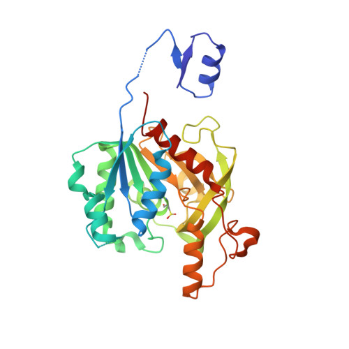
    A p-isothiocyanate phenylalanine mutant of the homodimeric protein homoserine o-succinyltransferase (MetA) was isolated in a temperature dependent selection from a library of metA mutants containing noncanonical amino acids. This mutant protein has a dramatic increase of 24 °C in thermal stability compared to the wild type protein. Peptide mapping experiments revealed that the isothiocyanate group forms a thiourea cross-link to the N terminal proline of the other monomer, despite the two positions being >30 Å apart in the X-ray crystal structure of the wild type protein. These results show that an expanded set of building blocks beyond the canonical 20 amino acids can lead to significant changes in the properties of proteins.


  • Organizational Affiliation
    • Department of Chemistry and Skaggs Institute for Chemical Biology , The Scripps Research Institute , La Jolla , California 92037 , United States.

Macromolecule Content 

  • Total Structure Weight: 70.7 kDa 
  • Atom Count: 5,388 
  • Modeled Residue Count: 583 
  • Deposited Residue Count: 604 
  • Unique protein chains: 1

Macromolecules

Find similar proteins by:|  3D Structure
Entity ID: 1
MoleculeChains  Sequence LengthOrganismDetailsImage
Homoserine O-succinyltransferase
A, B
302Escherichia coli K-12Mutation(s): 1 
Gene Names: metASmetAb4013JW3973
EC: 2.3.1.46
UniProt
Find proteins for P07623 (Escherichia coli (strain K12))
Explore P07623 
Go to UniProtKB:  P07623
Entity Groups
Sequence Clusters30% Identity50% Identity70% Identity90% Identity95% Identity100% Identity
UniProt GroupP07623
Sequence Annotations
Expand
Reference Sequence

Small Molecules

Ligands 3 Unique
IDChains Name / Formula / InChI Key2D Diagram3D Interactions
PEG

Query on PEG


Download:Ideal Coordinates CCD File
C [auth A],
D [auth A]
DI(HYDROXYETHYL)ETHER
C4 H10 O3
MTHSVFCYNBDYFN-UHFFFAOYSA-N
GOL

Query on GOL


Download:Ideal Coordinates CCD File
G [auth B]GLYCEROL
C3 H8 O3
PEDCQBHIVMGVHV-UHFFFAOYSA-N
FMT

Query on FMT


Download:Ideal Coordinates CCD File
E [auth A],
F [auth A],
H [auth B],
I [auth B],
J [auth B]
FORMIC ACID
C H2 O2
BDAGIHXWWSANSR-UHFFFAOYSA-N
Modified Residues  1 Unique
IDChains TypeFormula2D DiagramParent
CSD
Query on CSD
A, B
L-PEPTIDE LINKINGC3 H7 N O4 SCYS

Experimental Data & Validation

Experimental Data

  • Method: X-RAY DIFFRACTION
  • Resolution: 1.85 Å
  • R-Value Free:  0.218 (Depositor), 0.222 (DCC) 
  • R-Value Work:  0.169 (Depositor), 0.179 (DCC) 
  • R-Value Observed: 0.172 (Depositor) 
Space Group: P 21 21 21
Unit Cell:
Length ( Å )Angle ( ˚ )
a = 51.948α = 90
b = 100.036β = 90
c = 125.218γ = 90
Software Package:
Software NamePurpose
REFMACrefinement
XDSdata reduction
SCALAdata scaling
PHASERphasing
PDB_EXTRACTdata extraction

Structure Validation

View Full Validation Report


Entry History 

& Funding Information

Deposition Data


Funding OrganizationLocationGrant Number
National Institutes of Health/National Institute of General Medical Sciences (NIH/NIGMS)United StatesGM062159

Revision History  (Full details and data files)

  • Version 1.0: 2019-06-26
    Type: Initial release
  • Version 1.1: 2019-07-10
    Changes: Data collection, Database references
  • Version 1.2: 2020-01-01
    Changes: Author supporting evidence
  • Version 1.3: 2023-10-11
    Changes: Data collection, Database references, Refinement description
  • Version 1.4: 2024-11-06
    Changes: Structure summary