6MP1

Crystal structures of the murine class I major histocompatibility complex H-2Db in complex with the mutant TRP1-K8 peptide


Experimental Data Snapshot

  • Method: X-RAY DIFFRACTION
  • Resolution: 2.21 Å
  • R-Value Free: 0.244 
  • R-Value Work: 0.209 
  • R-Value Observed: 0.211 

wwPDB Validation   3D Report Full Report

Currently 6MP1 does not have a validation slider image.


This is version 1.5 of the entry. See complete history


Literature

Altered Binding of Tumor Antigenic Peptides to MHC Class I Affects CD8+T Cell-Effector Responses.

Clancy-Thompson, E.Devlin, C.A.Tyler, P.M.Servos, M.M.Ali, L.R.Ventre, K.S.Bhuiyan, M.A.Bruck, P.T.Birnbaum, M.E.Dougan, S.K.

(2018) Cancer Immunol Res 6: 1524-1536

  • DOI: https://doi.org/10.1158/2326-6066.CIR-18-0348
  • Primary Citation of Related Structures:  
    6MP0, 6MP1

  • PubMed Abstract: 

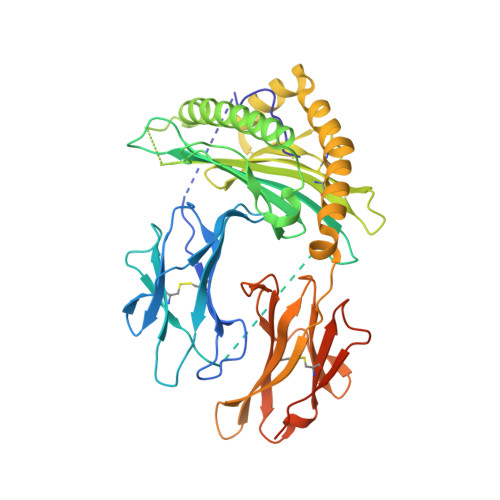
    T-cell priming occurs when a naïve T cell recognizes cognate peptide-MHC complexes on an activated antigen-presenting cell. The circumstances of this initial priming have ramifications on the fate of the newly primed T cell. Newly primed CD8 + T cells can embark onto different trajectories, with some becoming short-lived effector cells and others adopting a tissue resident or memory cell fate. To determine whether T-cell priming influences the quality of the effector T-cell response to tumors, we used transnuclear CD8 + T cells that recognize the melanoma antigen TRP1 using TRP1 high or TRP1 low TCRs that differ in both affinity and fine specificity. From a series of altered peptide ligands, we identified a point mutation (K8) in a nonanchor residue that, when analyzed crystallographically and biophysically, destabilized the peptide interaction with the MHC binding groove. In vitro , the K8 peptide induced robust proliferation of both TRP1 high and TRP1 low CD8 + T cells but did not induce expression of PD-1. Cytokine production from K8-stimulated TRP1 cells was minimal, whereas cytotoxicity was increased. Upon transfer into B16 tumor-bearing mice, the reference peptide (TRP1-M9)- and K8-stimulated TRP1 cells were equally effective at controlling tumor growth but accomplished this through different mechanisms. TRP1-M9-stimulated cells produced more IFNγ, whereas K8-stimulated cells accumulated to higher numbers and were more cytotoxic. We, therefore, conclude that TCR recognition of weakly binding peptides during priming can skew the effector function of tumor-specific CD8 + T cells.


  • Organizational Affiliation

    Department of Cancer Immunology and Virology, Dana-Farber Cancer Institute, Boston, Massachusetts.


Macromolecules
Find similar proteins by:  (by identity cutoff)  |  3D Structure
Entity ID: 1
MoleculeChains Sequence LengthOrganismDetailsImage
TRP1-K8 peptide, Beta-2-microglobulin,H-2 class I histocompatibility antigen, D-B alpha chain, chimeric construct447Mus musculusMutation(s): 3 
Gene Names: Tyrp1Tyrp-1B2mH2-D1
EC: 1.14.18
UniProt & NIH Common Fund Data Resources
Find proteins for P01899 (Mus musculus)
Explore P01899 
Go to UniProtKB:  P01899
Find proteins for P01887 (Mus musculus)
Explore P01887 
Go to UniProtKB:  P01887
IMPC:  MGI:88127
Find proteins for P07147 (Mus musculus)
Explore P07147 
Go to UniProtKB:  P07147
Entity Groups  
Sequence Clusters30% Identity50% Identity70% Identity90% Identity95% Identity100% Identity
UniProt GroupsP01899P01887P07147
Sequence Annotations
Expand
  • Reference Sequence
Small Molecules
Ligands 1 Unique
IDChains Name / Formula / InChI Key2D Diagram3D Interactions
NAG
Query on NAG

Download Ideal Coordinates CCD File 
B [auth A],
C [auth A]
2-acetamido-2-deoxy-beta-D-glucopyranose
C8 H15 N O6
OVRNDRQMDRJTHS-FMDGEEDCSA-N
Experimental Data & Validation

Experimental Data

  • Method: X-RAY DIFFRACTION
  • Resolution: 2.21 Å
  • R-Value Free: 0.244 
  • R-Value Work: 0.209 
  • R-Value Observed: 0.211 
  • Space Group: P 21 21 21
Unit Cell:
Length ( Å )Angle ( ˚ )
a = 52.569α = 90
b = 68.329β = 90
c = 117.787γ = 90
Software Package:
Software NamePurpose
PHENIXrefinement
XDSdata reduction
XSCALEdata scaling
PHASERphasing

Structure Validation

View Full Validation Report

Currently 6MP1 does not have a validation slider image.


Entry History & Funding Information

Deposition Data


Funding OrganizationLocationGrant Number
National Institutes of Health/National Cancer Institute (NIH/NCI)United StatesP30-CA14051
National Institutes of Health/National Cancer Institute (NIH/NCI)United StatesT32CA207021

Revision History  (Full details and data files)

  • Version 1.0: 2018-11-07
    Type: Initial release
  • Version 1.1: 2018-11-28
    Changes: Data collection, Database references
  • Version 1.2: 2018-12-12
    Changes: Data collection, Database references
  • Version 1.3: 2019-12-04
    Changes: Author supporting evidence
  • Version 1.4: 2020-07-29
    Type: Remediation
    Reason: Carbohydrate remediation
    Changes: Data collection, Derived calculations, Structure summary
  • Version 1.5: 2023-10-11
    Changes: Data collection, Database references, Refinement description, Structure summary