6MGH

X-ray structure of monomeric near-infrared fluorescent protein miRFP670nano


Experimental Data Snapshot

  • Method: X-RAY DIFFRACTION
  • Resolution: 1.95 Å
  • R-Value Free: 0.239 
  • R-Value Work: 0.182 
  • R-Value Observed: 0.183 

wwPDB Validation   3D Report Full Report


Ligand Structure Quality Assessment 


This is version 1.1 of the entry. See complete history


Literature

Smallest near-infrared fluorescent protein evolved from cyanobacteriochrome as versatile tag for spectral multiplexing.

Oliinyk, O.S.Shemetov, A.A.Pletnev, S.Shcherbakova, D.M.Verkhusha, V.V.

(2019) Nat Commun 10: 279-279

  • DOI: https://doi.org/10.1038/s41467-018-08050-8
  • Primary Citation of Related Structures:  
    6MGH

  • PubMed Abstract: 

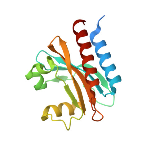
    From a single domain of cyanobacteriochrome (CBCR) we developed a near-infrared (NIR) fluorescent protein (FP), termed miRFP670nano, with excitation at 645 nm and emission at 670 nm. This is the first CBCR-derived NIR FP evolved to efficiently bind endogenous biliverdin chromophore and brightly fluoresce in mammalian cells. miRFP670nano is a monomer with molecular weight of 17 kDa that is 2-fold smaller than bacterial phytochrome (BphP)-based NIR FPs and 1.6-fold smaller than GFP-like FPs. Crystal structure of the CBCR-based NIR FP with biliverdin reveals a molecular basis of its spectral and biochemical properties. Unlike BphP-derived NIR FPs, miRFP670nano is highly stable to denaturation and degradation and can be used as an internal protein tag. miRFP670nano is an effective FRET donor for red-shifted NIR FPs, enabling engineering NIR FRET biosensors spectrally compatible with GFP-like FPs and blue-green optogenetic tools. miRFP670nano unlocks a new source of diverse CBCR templates for NIR FPs.


  • Organizational Affiliation

    Medicum, Faculty of Medicine, University of Helsinki, 00290, Helsinki, Finland.


Macromolecules
Find similar proteins by:  (by identity cutoff)  |  3D Structure
Entity ID: 1
MoleculeChains Sequence LengthOrganismDetailsImage
miRFP670nano
A, B, C, D, E
A, B, C, D, E, F, G, H
163Acaryochloris marinaMutation(s): 0 
Entity Groups  
Sequence Clusters30% Identity50% Identity70% Identity90% Identity95% Identity100% Identity
Sequence Annotations
Expand
  • Reference Sequence
Small Molecules
Ligands 3 Unique
IDChains Name / Formula / InChI Key2D Diagram3D Interactions
JRA
Query on JRA

Download Ideal Coordinates CCD File 
I [auth A]
J [auth B]
M [auth C]
P [auth D]
Q [auth E]
I [auth A],
J [auth B],
M [auth C],
P [auth D],
Q [auth E],
R [auth F],
S [auth G],
U [auth H]
3-[2-[(~{Z})-[5-[(~{Z})-[(3~{S},4~{R})-3-ethenyl-4-methyl-5-oxidanylidene-pyrrolidin-2-ylidene]methyl]-3-(3-hydroxy-3-oxopropyl)-4-methyl-pyrrol-2-ylidene]methyl]-5-[(~{Z})-(4-ethenyl-3-methyl-5-oxidanylidene-pyrrol-2-ylidene)methyl]-4-methyl-1~{H}-pyrrol-3-yl]propanoic acid
C33 H36 N4 O6
WLDQKQLRZDEERT-YTUMONHESA-N
GOL
Query on GOL

Download Ideal Coordinates CCD File 
K [auth B],
N [auth C],
T [auth G],
V [auth H]
GLYCEROL
C3 H8 O3
PEDCQBHIVMGVHV-UHFFFAOYSA-N
IPA
Query on IPA

Download Ideal Coordinates CCD File 
L [auth B],
O [auth C]
ISOPROPYL ALCOHOL
C3 H8 O
KFZMGEQAYNKOFK-UHFFFAOYSA-N
Experimental Data & Validation

Experimental Data

  • Method: X-RAY DIFFRACTION
  • Resolution: 1.95 Å
  • R-Value Free: 0.239 
  • R-Value Work: 0.182 
  • R-Value Observed: 0.183 
  • Space Group: C 1 2 1
Unit Cell:
Length ( Å )Angle ( ˚ )
a = 187.598α = 90
b = 72.124β = 129.99
c = 136.698γ = 90
Software Package:
Software NamePurpose
SCALEPACKdata scaling
REFMACrefinement
PDB_EXTRACTdata extraction
HKL-2000data reduction
MOLREPphasing

Structure Validation

View Full Validation Report


Ligand Structure Quality Assessment 


Entry History 

Deposition Data

  • Released Date: 2018-12-19 
  • Deposition Author(s): Pletnev, S.

Revision History  (Full details and data files)

  • Version 1.0: 2018-12-19
    Type: Initial release
  • Version 1.1: 2019-01-30
    Changes: Data collection, Database references