6MGB

Thermosulfurimonas dismutans KpsC, beta Kdo 2,4 transferase


Experimental Data Snapshot

  • Method: X-RAY DIFFRACTION
  • Resolution: 1.80 Å
  • R-Value Free: 0.213 
  • R-Value Work: 0.183 
  • R-Value Observed: 0.184 

wwPDB Validation   3D Report Full Report


Ligand Structure Quality Assessment 


This is version 1.6 of the entry. See complete history


Literature

Biosynthesis of a conserved glycolipid anchor for Gram-negative bacterial capsules.

Doyle, L.Ovchinnikova, O.G.Myler, K.Mallette, E.Huang, B.S.Lowary, T.L.Kimber, M.S.Whitfield, C.

(2019) Nat Chem Biol 15: 632-640

  • DOI: https://doi.org/10.1038/s41589-019-0276-8
  • Primary Citation of Related Structures:  
    6MGB, 6MGC, 6MGD

  • PubMed Abstract: 

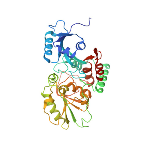
    Several important Gram-negative bacterial pathogens possess surface capsular layers composed of hypervariable long-chain polysaccharides linked via a conserved 3-deoxy-β-D-manno-oct-2-ulosonic acid (β-Kdo) oligosaccharide to a phosphatidylglycerol residue. The pathway for synthesis of the terminal glycolipid was elucidated by determining the structures of reaction intermediates. In Escherichia coli, KpsS transfers a single Kdo residue to phosphatidylglycerol; this primer is extended using a single enzyme (KpsC), possessing two cytidine 5'-monophosphate (CMP)-Kdo-dependent glycosyltransferase catalytic centers with different linkage specificities. The structure of the N-terminal β-(2→4) Kdo transferase from KpsC reveals two α/β domains, supplemented by several helices. The N-terminal Rossmann-like domain, typically responsible for acceptor binding, is severely reduced in size compared with canonical GT-B folds in glycosyltransferases. The similar structure of the C-terminal β-(2→7) Kdo transferase indicates a past gene duplication event. Both Kdo transferases have a narrow active site tunnel, lined with key residues shared with GT99 β-Kdo transferases. This enzyme provides the prototype for the GT107 family.


  • Organizational Affiliation

    Department of Molecular and Cellular Biology, University of Guelph, Guelph, Ontario, Canada.


Macromolecules
Find similar proteins by:  (by identity cutoff)  |  3D Structure
Entity ID: 1
MoleculeChains Sequence LengthOrganismDetailsImage
Capsular polysaccharide export system protein KpsC326Thermosulfurimonas dismutansMutation(s): 0 
Gene Names: TDIS_2046
UniProt
Find proteins for A0A179D1D1 (Thermosulfurimonas dismutans)
Explore A0A179D1D1 
Go to UniProtKB:  A0A179D1D1
Entity Groups  
Sequence Clusters30% Identity50% Identity70% Identity90% Identity95% Identity100% Identity
UniProt GroupA0A179D1D1
Sequence Annotations
Expand
  • Reference Sequence
Experimental Data & Validation

Experimental Data

  • Method: X-RAY DIFFRACTION
  • Resolution: 1.80 Å
  • R-Value Free: 0.213 
  • R-Value Work: 0.183 
  • R-Value Observed: 0.184 
  • Space Group: P 21 21 21
Unit Cell:
Length ( Å )Angle ( ˚ )
a = 50.02α = 90
b = 65.54β = 90
c = 121.08γ = 90
Software Package:
Software NamePurpose
PHENIXrefinement
XDSdata reduction
XSCALEdata scaling
PHENIXphasing

Structure Validation

View Full Validation Report


Ligand Structure Quality Assessment 


Entry History & Funding Information

Deposition Data


Funding OrganizationLocationGrant Number
Natural Sciences and Engineering Research Council (NSERC, Canada)Canada04045-2015

Revision History  (Full details and data files)

  • Version 1.0: 2019-03-27
    Type: Initial release
  • Version 1.1: 2019-05-01
    Changes: Data collection, Database references
  • Version 1.2: 2019-05-08
    Changes: Data collection, Database references
  • Version 1.3: 2019-05-15
    Changes: Data collection, Database references
  • Version 1.4: 2019-05-29
    Changes: Data collection, Database references
  • Version 1.5: 2020-01-08
    Changes: Author supporting evidence
  • Version 1.6: 2024-03-13
    Changes: Data collection, Database references, Derived calculations