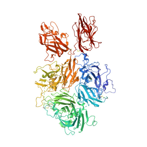
6MF0
Crystal Structure Determination of Human/Porcine Chimera Coagulation Factor VIII
- PDB DOI: https://doi.org/10.2210/pdb6MF0/pdb
- Classification: BLOOD CLOTTING
- Organism(s): Sus scrofa, Homo sapiens
- Expression System: Cricetulus griseus
- Mutation(s): No 
- Membrane Protein: Yes  OPM
- Deposited: 2018-09-07 Released: 2019-09-11 
- Funding Organization(s): National Institutes of Health/National Heart, Lung, and Blood Institute (NIH/NHLBI), National Science Foundation (NSF, United States)
Experimental Data Snapshot
- Method: X-RAY DIFFRACTION
- Resolution: 3.20 Å
- R-Value Free: 0.287 
- R-Value Work: 0.206 
- R-Value Observed: 0.210 
wwPDB Validation   3D Report Full Report
Currently 6MF0 does not have a validation slider image.
This is version 2.1 of the entry. See complete history. 
Macromolecules
Find similar proteins by:
(by identity cutoff) | 3D Structure
Entity ID: 1 | |||||
---|---|---|---|---|---|
Molecule | Chains | Sequence Length | Organism | Details | Image |
Coagulation factor VIII chimera | 1,467 | Sus scrofa, Homo sapiens | Mutation(s): 0  Gene Names: F8, CF8, F8C Membrane Entity: Yes  | ||
UniProt & NIH Common Fund Data Resources | |||||
Find proteins for P12263 (Sus scrofa) Explore P12263  Go to UniProtKB:  P12263 | |||||
Find proteins for P00451 (Homo sapiens) Explore P00451  Go to UniProtKB:  P00451 | |||||
PHAROS:  P00451 GTEx:  ENSG00000185010  | |||||
Entity Groups   | |||||
Sequence Clusters | 30% Identity50% Identity70% Identity90% Identity95% Identity100% Identity | ||||
UniProt Groups | P12263P00451 | ||||
Sequence AnnotationsExpand | |||||
|
Oligosaccharides
Entity ID: 2 | |||||
---|---|---|---|---|---|
Molecule | Chains | Length | 2D Diagram | Glycosylation | 3D Interactions |
alpha-D-mannopyranose-(1-3)-[alpha-D-mannopyranose-(1-6)]beta-D-mannopyranose-(1-4)-2-acetamido-2-deoxy-beta-D-glucopyranose-(1-4)-2-acetamido-2-deoxy-beta-D-glucopyranose | C, E, F | 5 | N-Glycosylation | ||
Glycosylation Resources | |||||
GlyTouCan:  G22768VO GlyCosmos:  G22768VO GlyGen:  G22768VO |
Entity ID: 3 | |||||
---|---|---|---|---|---|
Molecule | Chains | Length | 2D Diagram | Glycosylation | 3D Interactions |
beta-D-mannopyranose-(1-4)-2-acetamido-2-deoxy-beta-D-glucopyranose-(1-4)-2-acetamido-2-deoxy-beta-D-glucopyranose | D | 3 | N-Glycosylation | ||
Glycosylation Resources | |||||
GlyTouCan:  G15407YE GlyCosmos:  G15407YE GlyGen:  G15407YE |
Entity ID: 4 | |||||
---|---|---|---|---|---|
Molecule | Chains | Length | 2D Diagram | Glycosylation | 3D Interactions |
2-acetamido-2-deoxy-beta-D-glucopyranose-(1-4)-alpha-D-mannopyranose-(1-6)-[alpha-D-mannopyranose-(1-3)]beta-D-mannopyranose-(1-4)-2-acetamido-2-deoxy-beta-D-glucopyranose-(1-4)-[alpha-L-fucopyranose-(1-6)]2-acetamido-2-deoxy-beta-D-glucopyranose | G | 7 | N-Glycosylation | ||
Glycosylation Resources | |||||
GlyTouCan:  G35459KZ GlyCosmos:  G35459KZ GlyGen:  G35459KZ |
Entity ID: 5 | |||||
---|---|---|---|---|---|
Molecule | Chains | Length | 2D Diagram | Glycosylation | 3D Interactions |
alpha-D-mannopyranose-(1-2)-alpha-D-mannopyranose-(1-3)-[alpha-D-mannopyranose-(1-6)]beta-D-mannopyranose-(1-4)-2-acetamido-2-deoxy-beta-D-glucopyranose-(1-4)-2-acetamido-2-deoxy-beta-D-glucopyranose | H | 6 | N-Glycosylation | ||
Glycosylation Resources | |||||
GlyTouCan:  G56014GC GlyCosmos:  G56014GC GlyGen:  G56014GC |
Small Molecules
Ligands 3 Unique | |||||
---|---|---|---|---|---|
ID | Chains | Name / Formula / InChI Key | 2D Diagram | 3D Interactions | |
ZN Query on ZN | J [auth A], M [auth B] | ZINC ION Zn PTFCDOFLOPIGGS-UHFFFAOYSA-N | |||
CU1 Query on CU1 | K [auth A], N [auth B] | COPPER (I) ION Cu VMQMZMRVKUZKQL-UHFFFAOYSA-N | |||
CA Query on CA | I [auth A], L [auth B] | CALCIUM ION Ca BHPQYMZQTOCNFJ-UHFFFAOYSA-N |
Experimental Data & Validation
Experimental Data
- Method: X-RAY DIFFRACTION
- Resolution: 3.20 Å
- R-Value Free: 0.287 
- R-Value Work: 0.206 
- R-Value Observed: 0.210 
- Space Group: P 1 21 1
Unit Cell:
Length ( Å ) | Angle ( ˚ ) |
---|---|
a = 72.005 | α = 90 |
b = 135.863 | β = 90.151 |
c = 196.107 | γ = 90 |
Software Name | Purpose |
---|---|
PHENIX | refinement |
HKL-2000 | data reduction |
HKL-2000 | data scaling |
PHASER | phasing |
Structure Validation
View Full Validation Report
Currently 6MF0 does not have a validation slider image.
Entry History & Funding Information
Deposition Data
- Released Date: 2019-09-11  Deposition Author(s): Smith, I.W., Spiegel, P.C.
Funding Organization | Location | Grant Number |
---|---|---|
National Institutes of Health/National Heart, Lung, and Blood Institute (NIH/NHLBI) | United States | R15 HL103518 |
National Science Foundation (NSF, United States) | United States | MRI 1429164 |
Revision History (Full details and data files)
- Version 1.0: 2019-09-11
Type: Initial release - Version 1.1: 2019-11-27
Changes: Author supporting evidence, Data collection - Version 1.2: 2020-01-15
Changes: Database references - Version 2.0: 2020-07-29
Type: Remediation
Reason: Carbohydrate remediation
Changes: Advisory, Atomic model, Data collection, Derived calculations, Structure summary - Version 2.1: 2023-10-11
Changes: Data collection, Database references, Derived calculations, Refinement description, Structure summary