6MBC | pdb_00006mbc

Human Bfl-1 in complex with the designed peptide dF4


Experimental Data Snapshot

  • Method: X-RAY DIFFRACTION
  • Resolution: 1.75 Å
  • R-Value Free: 
    0.212 (Depositor), 0.215 (DCC) 
  • R-Value Work: 
    0.183 (Depositor), 0.187 (DCC) 
  • R-Value Observed: 
    0.186 (Depositor) 

wwPDB Validation 3D Report Full Report

Validation slider image for 6MBC

This is version 1.3 of the entry. See complete history

Literature

Tertiary Structural Motif Sequence Statistics Enable Facile Prediction and Design of Peptides that Bind Anti-apoptotic Bfl-1 and Mcl-1.

Frappier, V.Jenson, J.M.Zhou, J.Grigoryan, G.Keating, A.E.

(2019) Structure 27: 606-617.e5

  • DOI: https://doi.org/10.1016/j.str.2019.01.008
  • Primary Citation Related Structures: 
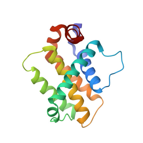
    6MBB, 6MBC, 6MBD, 6MBE

  • PubMed Abstract: 

    Understanding the relationship between protein sequence and structure well enough to design new proteins with desired functions is a longstanding goal in protein science. Here, we show that recurring tertiary structural motifs (TERMs) in the PDB provide rich information for protein-peptide interaction prediction and design. TERM statistics can be used to predict peptide binding energies for Bcl-2 family proteins as accurately as widely used structure-based tools. Furthermore, design using TERM energies (dTERMen) rapidly and reliably generates high-affinity peptide binders of anti-apoptotic proteins Bfl-1 and Mcl-1 with just 15%-38% sequence identity to any known native Bcl-2 family protein ligand. High-resolution structures of four designed peptides bound to their targets provide opportunities to analyze the strengths and limitations of the computational design method. Our results support dTERMen as a powerful approach that can complement existing tools for protein engineering.


  • Organizational Affiliation
    • Department of Biology, Massachusetts Institute of Technology, Cambridge, MA 02139, USA.

Macromolecule Content 

  • Total Structure Weight: 20.26 kDa 
  • Atom Count: 1,541 
  • Modeled Residue Count: 176 
  • Deposited Residue Count: 177 
  • Unique protein chains: 2

Macromolecules

Find similar proteins by:|  3D Structure
Entity ID: 1
MoleculeChains  Sequence LengthOrganismDetailsImage
Bcl-2-related protein A1152Homo sapiensMutation(s): 0 
Gene Names: BCL2A1BCL2L5BFL1GRSHBPA1
UniProt & NIH Common Fund Data Resources
Find proteins for Q16548 (Homo sapiens)
Explore Q16548 
Go to UniProtKB:  Q16548
PHAROS:  Q16548
GTEx:  ENSG00000140379 
Entity Groups
Sequence Clusters30% Identity50% Identity70% Identity90% Identity95% Identity100% Identity
UniProt GroupQ16548
Sequence Annotations
Expand
Reference Sequence
Find similar proteins by:  Sequence   |   3D Structure  
Entity ID: 2
MoleculeChains  Sequence LengthOrganismDetailsImage
dF425synthetic constructMutation(s): 0 

Experimental Data & Validation

Experimental Data

  • Method: X-RAY DIFFRACTION
  • Resolution: 1.75 Å
  • R-Value Free:  0.212 (Depositor), 0.215 (DCC) 
  • R-Value Work:  0.183 (Depositor), 0.187 (DCC) 
  • R-Value Observed: 0.186 (Depositor) 
Space Group: P 1 21 1
Unit Cell:
Length ( Å )Angle ( ˚ )
a = 43.466α = 90
b = 42.905β = 114.21
c = 46.666γ = 90
Software Package:
Software NamePurpose
HKL-2000data reduction
HKL-2000data scaling
PHENIXrefinement
PDB_EXTRACTdata extraction
PHENIXphasing

Structure Validation

View Full Validation Report


Entry History 

& Funding Information

Deposition Data


Funding OrganizationLocationGrant Number
National Institutes of Health/National Institute of General Medical Sciences (NIH/NIGMS)United StatesR01-GM110048

Revision History  (Full details and data files)

  • Version 1.0: 2019-03-06
    Type: Initial release
  • Version 1.1: 2019-04-10
    Changes: Data collection, Database references
  • Version 1.2: 2020-01-01
    Changes: Author supporting evidence
  • Version 1.3: 2024-10-23
    Changes: Data collection, Database references, Structure summary