6MA8

Human CYP3A4 bound to PMSF


Experimental Data Snapshot

  • Method: X-RAY DIFFRACTION
  • Resolution: 1.83 Å
  • R-Value Free: 0.229 
  • R-Value Work: 0.182 
  • R-Value Observed: 0.185 

wwPDB Validation   3D Report Full Report


Ligand Structure Quality Assessment 


This is version 1.3 of the entry. See complete history


Literature

Interaction of Human Drug-Metabolizing CYP3A4 with Small Inhibitory Molecules.

Sevrioukova, I.

(2019) Biochemistry 58: 930-939

  • DOI: https://doi.org/10.1021/acs.biochem.8b01221
  • Primary Citation of Related Structures:  
    6MA6, 6MA7, 6MA8

  • PubMed Abstract: 

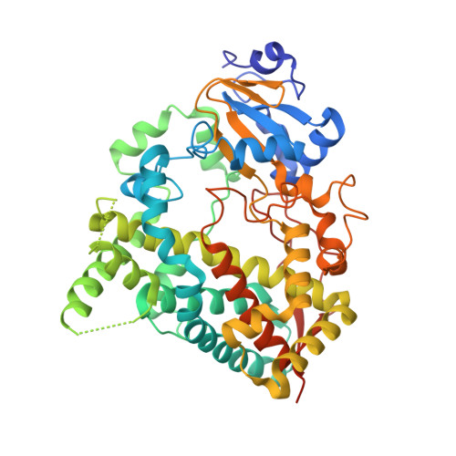
    Binding of small inhibitory compounds to human cytochrome P450 3A4 (CYP3A4) could interfere with drug metabolism and lead to drug-drug interactions, the underlying mechanism of which is not fully understood due to insufficient structural information. This study investigated the interaction of recombinant CYP3A4 with a nonspecific inhibitor metyrapone, antifungal drug fluconazole, and protease inhibitor phenylmethanesulfonyl fluoride (PMSF). Metyrapone and fluconazole are classic type II ligands that inhibit CYP3A4 with medium strength by ligating to the heme iron, whereas PMSF, lacking the heme-ligating moiety, acts as a weak type I ligand and inhibitor of CYP3A4. High-resolution crystal structures revealed that the orientation of metyrapone is similar but not identical to that in the previously reported 1W0G model, whereas the flexible fluconazole adapts a conformer markedly different from that observed in the target CYP51 enzymes, which could explain its high potential for cross-reactivity. Besides hydrophobic and aromatic interactions with the heme and active site residues, both drugs establish water-mediated contacts that stabilize the inhibitory complexes. PMSF also binds near the catalytic center, with the phenyl group parallel to the heme. However, it does not displace the water ligand and is held in place via strong H-bonds formed by the sulfofluoride moiety with Ser119 and Arg212. Collectively, our data suggest that PMSF might have multiple binding sites and likely occupies the high-affinity site in the crystal structure. Moreover, its hydrolysis product, phenylmethanesulfonic acid, can also access and be retained in the CYP3A4 active site. Therefore, to avoid experimental artifacts, PMSF should be excluded from purification and assay solutions.


  • Organizational Affiliation

    Department of Molecular Biology and Biochemistry , University of California , Irvine , California 92697-3900 , United States.


Macromolecules
Find similar proteins by:  (by identity cutoff)  |  3D Structure
Entity ID: 1
MoleculeChains Sequence LengthOrganismDetailsImage
Cytochrome P450 3A4487Homo sapiensMutation(s): 0 
Gene Names: CYP3A4CYP3A3
EC: 1.14.14 (PDB Primary Data), 1.14.14.56 (PDB Primary Data), 1.14.14.55 (PDB Primary Data)
UniProt & NIH Common Fund Data Resources
Find proteins for P08684 (Homo sapiens)
Explore P08684 
Go to UniProtKB:  P08684
PHAROS:  P08684
GTEx:  ENSG00000160868 
Entity Groups  
Sequence Clusters30% Identity50% Identity70% Identity90% Identity95% Identity100% Identity
UniProt GroupP08684
Sequence Annotations
Expand
  • Reference Sequence
Small Molecules
Ligands 5 Unique
IDChains Name / Formula / InChI Key2D Diagram3D Interactions
HEM
Query on HEM

Download Ideal Coordinates CCD File 
B [auth A]PROTOPORPHYRIN IX CONTAINING FE
C34 H32 Fe N4 O4
KABFMIBPWCXCRK-RGGAHWMASA-L
PMF
Query on PMF

Download Ideal Coordinates CCD File 
E [auth A]PHENYLMETHYLSULFONYL FLUORIDE
C7 H7 F O2 S
YBYRMVIVWMBXKQ-UHFFFAOYSA-N
GOL
Query on GOL

Download Ideal Coordinates CCD File 
C [auth A],
D [auth A]
GLYCEROL
C3 H8 O3
PEDCQBHIVMGVHV-UHFFFAOYSA-N
DMS
Query on DMS

Download Ideal Coordinates CCD File 
M [auth A]DIMETHYL SULFOXIDE
C2 H6 O S
IAZDPXIOMUYVGZ-UHFFFAOYSA-N
EDO
Query on EDO

Download Ideal Coordinates CCD File 
F [auth A]
G [auth A]
H [auth A]
I [auth A]
J [auth A]
F [auth A],
G [auth A],
H [auth A],
I [auth A],
J [auth A],
K [auth A],
L [auth A]
1,2-ETHANEDIOL
C2 H6 O2
LYCAIKOWRPUZTN-UHFFFAOYSA-N
Binding Affinity Annotations 
IDSourceBinding Affinity
PMF Binding MOAD:  6MA8 Kd: 2.65e+6 (nM) from 1 assay(s)
Experimental Data & Validation

Experimental Data

  • Method: X-RAY DIFFRACTION
  • Resolution: 1.83 Å
  • R-Value Free: 0.229 
  • R-Value Work: 0.182 
  • R-Value Observed: 0.185 
  • Space Group: I 2 2 2
Unit Cell:
Length ( Å )Angle ( ˚ )
a = 77.22α = 90
b = 99.62β = 90
c = 131.88γ = 90
Software Package:
Software NamePurpose
PHENIXrefinement
MOSFLMdata reduction
SCALAdata scaling
PHASERphasing

Structure Validation

View Full Validation Report


Ligand Structure Quality Assessment 


Entry History & Funding Information

Deposition Data


Funding OrganizationLocationGrant Number
National Institutes of Health/National Institute of Environmental Health Sciences (NIH/NIEHS)United StatesES025767

Revision History  (Full details and data files)

  • Version 1.0: 2019-01-16
    Type: Initial release
  • Version 1.1: 2019-06-12
    Changes: Data collection, Database references
  • Version 1.2: 2019-12-18
    Changes: Author supporting evidence
  • Version 1.3: 2023-10-11
    Changes: Data collection, Database references, Refinement description