6M7L | pdb_00006m7l

Complex of OxyA with the X-domain from GPA biosynthesis


Experimental Data Snapshot

  • Method: X-RAY DIFFRACTION
  • Resolution: 2.65 Å
  • R-Value Free: 
    0.264 (Depositor), 0.237 (DCC) 
  • R-Value Work: 
    0.214 (Depositor) 
  • R-Value Observed: 
    0.217 (Depositor) 

Starting Model: experimental
View more details

wwPDB Validation 3D Report Full Report

Validation slider image for 6M7L

Ligand Structure Quality Assessment 


This is version 1.4 of the entry. See complete history

Literature

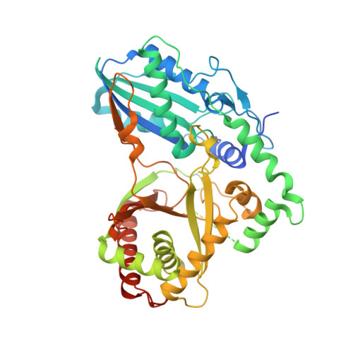
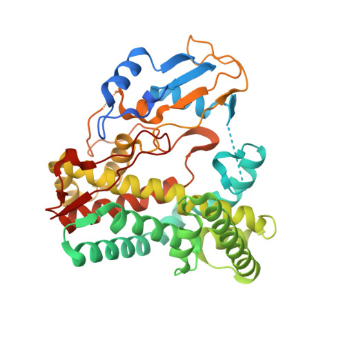
Kistamicin biosynthesis reveals the biosynthetic requirements for production of highly crosslinked glycopeptide antibiotics.

Greule, A.Izore, T.Iftime, D.Tailhades, J.Schoppet, M.Zhao, Y.Peschke, M.Ahmed, I.Kulik, A.Adamek, M.Goode, R.J.A.Schittenhelm, R.B.Kaczmarski, J.A.Jackson, C.J.Ziemert, N.Krenske, E.H.De Voss, J.J.Stegmann, E.Cryle, M.J.

(2019) Nat Commun 10: 2613-2613

  • DOI: https://doi.org/10.1038/s41467-019-10384-w
  • Primary Citation Related Structures: 
    6M7L

  • PubMed Abstract: 

    Kistamicin is a divergent member of the glycopeptide antibiotics, a structurally complex class of important, clinically relevant antibiotics often used as the last resort against resistant bacteria. The extensively crosslinked structure of these antibiotics that is essential for their activity makes their chemical synthesis highly challenging and limits their production to bacterial fermentation. Kistamicin contains three crosslinks, including an unusual 15-membered A-O-B ring, despite the presence of only two Cytochrome P450 Oxy enzymes thought to catalyse formation of such crosslinks within the biosynthetic gene cluster. In this study, we characterise the kistamicin cyclisation pathway, showing that the two Oxy enzymes are responsible for these crosslinks within kistamicin and that they function through interactions with the X-domain, unique to glycopeptide antibiotic biosynthesis. We also show that the kistamicin OxyC enzyme is a promiscuous biocatalyst, able to install multiple crosslinks into peptides containing phenolic amino acids.


  • Organizational Affiliation
    • Department of Biochemistry and Molecular Biology, The Monash Biomedicine Discovery Institute, Monash University, Clayton, VIC, 3800, Australia.

Macromolecule Content 

  • Total Structure Weight: 99.16 kDa 
  • Atom Count: 6,233 
  • Modeled Residue Count: 792 
  • Deposited Residue Count: 890 
  • Unique protein chains: 2

Macromolecules

Find similar proteins by:|  3D Structure
Entity ID: 1
MoleculeChains  Sequence LengthOrganismDetailsImage
Putative non-ribosomal peptide synthetaseA [auth B]486[Actinomadura] parvosata subsp. kistnaeMutation(s): 0 
Gene Names: KIS93_04814
Find similar proteins by:|  3D Structure
Entity ID: 2
MoleculeChains  Sequence LengthOrganismDetailsImage
Putative cytochrome P450 hydroxylaseB [auth A]404[Actinomadura] parvosata subsp. kistnaeMutation(s): 0 
Gene Names: KIS93_04812

Small Molecules

Ligands 1 Unique
IDChains Name / Formula / InChI Key2D Diagram3D Interactions
HEM

Query on HEM


Download:Ideal Coordinates CCD File
C [auth A]PROTOPORPHYRIN IX CONTAINING FE
C34 H32 Fe N4 O4
KABFMIBPWCXCRK-RGGAHWMASA-L

Experimental Data & Validation

Experimental Data

  • Method: X-RAY DIFFRACTION
  • Resolution: 2.65 Å
  • R-Value Free:  0.264 (Depositor), 0.237 (DCC) 
  • R-Value Work:  0.214 (Depositor) 
  • R-Value Observed: 0.217 (Depositor) 
Space Group: C 1 2 1
Unit Cell:
Length ( Å )Angle ( ˚ )
a = 118.834α = 90
b = 87.344β = 95.769
c = 95.955γ = 90
Software Package:
Software NamePurpose
PHENIXrefinement
XDSdata reduction
Aimlessdata scaling
PHENIXphasing

Structure Validation

View Full Validation Report


Ligand Structure Quality Assessment 


Entry History 

& Funding Information

Deposition Data


Funding OrganizationLocationGrant Number
National Health and Medical Research Council (NHMRC, Australia)AustraliaAPP1140619
Australian Research Council (ARC)AustraliaDP170102220
German Research Foundation (DFG)GermanyCR 392/1-1

Revision History  (Full details and data files)

  • Version 1.0: 2019-05-22
    Type: Initial release
  • Version 1.1: 2019-06-26
    Changes: Data collection, Database references
  • Version 1.2: 2019-09-04
    Changes: Data collection, Structure summary
  • Version 1.3: 2020-01-01
    Changes: Author supporting evidence
  • Version 1.4: 2023-10-11
    Changes: Data collection, Database references, Refinement description