6M1X

Crystal structure of Phosphoserine Phosphatase in complex with 3-Phosphoglyceric Acid from Entamoeba histolytica


Experimental Data Snapshot

  • Method: X-RAY DIFFRACTION
  • Resolution: 2.48 Å
  • R-Value Free: 0.256 
  • R-Value Work: 0.228 
  • R-Value Observed: 0.230 

wwPDB Validation   3D Report Full Report


Ligand Structure Quality Assessment 


This is version 1.2 of the entry. See complete history


Literature

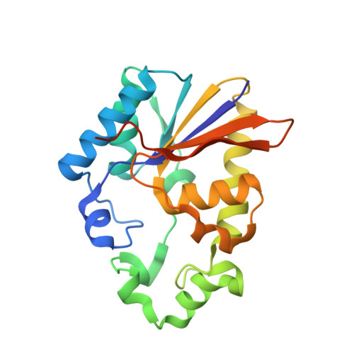
Structural analysis of EhPSP in complex with 3-phosphoglyceric acid from Entamoeba histolytica reveals a basis for its lack of phosphoglycerate mutase activity.

Kumari, P.Vijayan, R.Gourinath, S.

(2021) Int J Biol Macromol 178: 1-10

  • DOI: https://doi.org/10.1016/j.ijbiomac.2021.02.153
  • Primary Citation of Related Structures:  
    6M1X

  • PubMed Abstract: 

    Entamoeba histolytica phosphoserine phosphatase (EhPSP), a regulatory enzyme in the serine biosynthetic pathway, is also a structural homolog of cofactor-dependent phosphoglycerate mutase (dPGM). However, despite sharing many of its catalytic residues with dPGM, EhPSP displays no significant mutase activity. In the current work, we determined a crystal structure of EhPSP in complex with 3-PGA to 2.5 Å resolution and observed striking differences between the orientation of 3-PGA bound to EhPSP and that to its other homologous structures. We also performed computational modeling and simulations of the intermediate 2,3-bisphosphoglyceric acid into the active site of EhPSP to better understand its mechanistic details. Based on these results and those of a similar study with the dPGMs from E. coli and B. pseudomallei, the affinity of EhPSP for 2,3-BPG was concluded to be lower than those of the other proteins. Moreover, a different set of 2,3-BPG interacting residues was observed in EhPSP compared to dPGMs, with all of the crucial interacting residues of dPGMs either missing or substituted with weakly interacting residues. This study has expanded our understanding, at the structural level, of the inability of EhPSP to catalyze the mutase reaction and has strengthened earlier conclusions indicating it to be a true phosphatase.


  • Organizational Affiliation

    School of Life Sciences, Jawaharlal Nehru University, New Delhi, India.


Macromolecules
Find similar proteins by:  (by identity cutoff)  |  3D Structure
Entity ID: 1
MoleculeChains Sequence LengthOrganismDetailsImage
Phosphoglycerate mutase family protein
A, B, C, D
211Entamoeba histolyticaMutation(s): 0 
Gene Names: EHI_129820
UniProt
Find proteins for C4M5P9 (Entamoeba histolytica (strain ATCC 30459 / HM-1:IMSS / ABRM))
Explore C4M5P9 
Go to UniProtKB:  C4M5P9
Entity Groups  
Sequence Clusters30% Identity50% Identity70% Identity90% Identity95% Identity100% Identity
UniProt GroupC4M5P9
Sequence Annotations
Expand
  • Reference Sequence
Experimental Data & Validation

Experimental Data

  • Method: X-RAY DIFFRACTION
  • Resolution: 2.48 Å
  • R-Value Free: 0.256 
  • R-Value Work: 0.228 
  • R-Value Observed: 0.230 
  • Space Group: C 2 2 21
Unit Cell:
Length ( Å )Angle ( ˚ )
a = 96.976α = 90
b = 112.388β = 90
c = 177.428γ = 90
Software Package:
Software NamePurpose
REFMACrefinement
PDB_EXTRACTdata extraction
HKL-2000data reduction
HKL-2000data scaling
PHASERphasing

Structure Validation

View Full Validation Report


Ligand Structure Quality Assessment 


Entry History & Funding Information

Deposition Data


Funding OrganizationLocationGrant Number
Science and Engineering Research Board (SERB)--

Revision History  (Full details and data files)

  • Version 1.0: 2021-03-03
    Type: Initial release
  • Version 1.1: 2021-09-22
    Changes: Database references
  • Version 1.2: 2023-11-29
    Changes: Data collection, Refinement description