6LZO

Thermolysin with 1,10-phenanthroline

  • Classification: HYDROLASE
  • Organism(s): Bacillus thermoproteolyticus
  • Mutation(s): No 

  • Deposited: 2020-02-19 Released: 2021-01-27 
  • Deposition Author(s): Nam, K.H.
  • Funding Organization(s): National Research Foundation (NRF, Korea)

Experimental Data Snapshot

  • Method: X-RAY DIFFRACTION
  • Resolution: 1.80 Å
  • R-Value Free: 0.211 
  • R-Value Work: 0.172 
  • R-Value Observed: 0.175 

wwPDB Validation   3D Report Full Report


Ligand Structure Quality Assessment 


This is version 1.1 of the entry. See complete history


Literature

Structural analysis of metal chelation of the metalloproteinase thermolysin by 1,10-phenanthroline.

Nam, K.H.

(2021) J Inorg Biochem 215: 111319-111319

  • DOI: https://doi.org/10.1016/j.jinorgbio.2020.111319
  • Primary Citation of Related Structures:  
    6LZN, 6LZO

  • PubMed Abstract: 

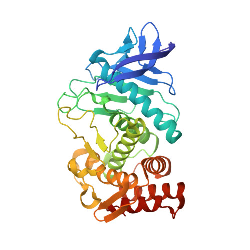
    Metalloproteases and their inhibitors are important in numerous fundamental biochemical phenomena and medical applications. The heterocyclic organic compound, 1,10-phenanthroline, forms a complex with transition metal ions and is a Zn 2+ -chelating metalloprotease inhibitor; however, the mechanism of 1,10-phenanthroline-based chelation inhibition has not been fully elucidated. This study aimed to understand the structural basis of zinc metalloproteinase inhibition by 1,10-phenanthroline. Herein, the crystal structure of thermolysin was determined in the absence and presence of 1,10-phenanthroline at 1.5 and 1.8 Å, respectively. In native thermolysin, Zn 2+ at the active site is tetrahedrally coordinated by His142, His146, Glu166, and water molecule and contains three Ca 2+ ions, which are involved in thermostability. In the crystal structure of 1,10-phenanthroline-treated thermolysin crystal, seven 1,10-phenanthroline molecules were observed on the surface of thermolysin. These molecules are stabilized by π- π stacking interactions with aromatic amino acids (Phe63, Tyr66, Tyr110, His216, and Try251) or between the 1,10-phenanthrolines. Moreover, interactions with Ser5 and Arg101 were also observed. In this structure, Zn 2+ at the active site was completely chelated, but no large conformational changes were observed in Zn 2+ coordination with amino acid residues. Ca 2+ at the Ca3 site exposed to the solvent was chelated by 1,10-phenanthroline, resulting in a conformational change in the side chain of Asp56 and Gln61. Based on the surface structure, for 1,10-phenanthroline to chelate a metal, it is important that the metal is exposed on the protein surface and that there is no steric hindrance impairing 1,10-phenanthroline access by the amino acids around the metal.


  • Organizational Affiliation

    Department of Life Science, Pohang University of Science and Technology, Pohang 37673, Republic of Korea. Electronic address: structures@postech.ac.kr.


Macromolecules
Find similar proteins by:  (by identity cutoff)  |  3D Structure
Entity ID: 1
MoleculeChains Sequence LengthOrganismDetailsImage
Thermolysin316Bacillus thermoproteolyticusMutation(s): 0 
EC: 3.4.24.27
UniProt
Find proteins for P00800 (Bacillus thermoproteolyticus)
Explore P00800 
Go to UniProtKB:  P00800
Entity Groups  
Sequence Clusters30% Identity50% Identity70% Identity90% Identity95% Identity100% Identity
UniProt GroupP00800
Sequence Annotations
Expand
  • Reference Sequence
Small Molecules
Ligands 2 Unique
IDChains Name / Formula / InChI Key2D Diagram3D Interactions
PHN (Subject of Investigation/LOI)
Query on PHN

Download Ideal Coordinates CCD File 
D [auth A]
E [auth A]
F [auth A]
G [auth A]
H [auth A]
D [auth A],
E [auth A],
F [auth A],
G [auth A],
H [auth A],
I [auth A],
J [auth A]
1,10-PHENANTHROLINE
C12 H8 N2
DGEZNRSVGBDHLK-UHFFFAOYSA-N
CA
Query on CA

Download Ideal Coordinates CCD File 
B [auth A],
C [auth A]
CALCIUM ION
Ca
BHPQYMZQTOCNFJ-UHFFFAOYSA-N
Experimental Data & Validation

Experimental Data

  • Method: X-RAY DIFFRACTION
  • Resolution: 1.80 Å
  • R-Value Free: 0.211 
  • R-Value Work: 0.172 
  • R-Value Observed: 0.175 
  • Space Group: P 61 2 2
Unit Cell:
Length ( Å )Angle ( ˚ )
a = 92.772α = 90
b = 92.772β = 90
c = 129.103γ = 120
Software Package:
Software NamePurpose
REFMACrefinement
HKL-2000data scaling
PDB_EXTRACTdata extraction
HKL-2000data reduction
MOLREPphasing

Structure Validation

View Full Validation Report


Ligand Structure Quality Assessment 


Entry History & Funding Information

Deposition Data

  • Released Date: 2021-01-27 
  • Deposition Author(s): Nam, K.H.

Funding OrganizationLocationGrant Number
National Research Foundation (NRF, Korea)Korea, Republic OfNRF-2017R1D1A1B03033087
National Research Foundation (NRF, Korea)Korea, Republic OfNRF-2017M3A9F6029736

Revision History  (Full details and data files)

  • Version 1.0: 2021-01-27
    Type: Initial release
  • Version 1.1: 2023-11-29
    Changes: Data collection, Database references, Refinement description