6LZ4

Crystal structure of PMab-1 Fv-clasp fragment with its antigen peptide


Experimental Data Snapshot

  • Method: X-RAY DIFFRACTION
  • Resolution: 2.49 Å
  • R-Value Free: 0.269 
  • R-Value Work: 0.245 
  • R-Value Observed: 0.246 

wwPDB Validation   3D Report Full Report


This is version 1.2 of the entry. See complete history


Literature

Site-specific epitope insertion into recombinant proteins using the MAP tag system.

Wakasa, A.Kaneko, M.K.Kato, Y.Takagi, J.Arimori, T.

(2020) J Biochem 168: 375-384

  • DOI: https://doi.org/10.1093/jb/mvaa054
  • Primary Citation of Related Structures:  
    6LZ4

  • PubMed Abstract: 

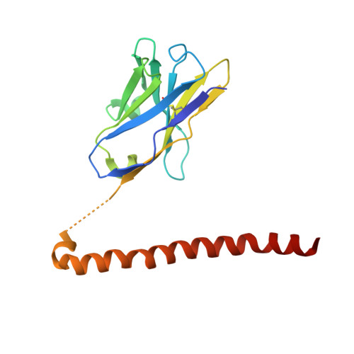
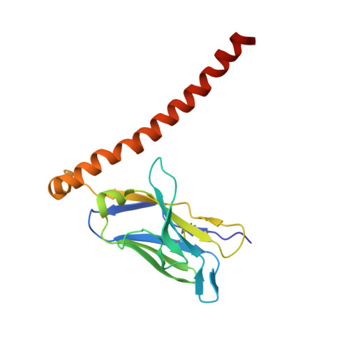
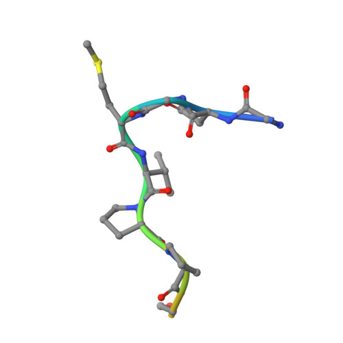
    The MAP tag system comprises a 14-residue peptide derived from mouse podoplanin and its high-affinity monoclonal antibody PMab-1. We determined the crystal structure of PMab-1 complexed with the MAP tag peptide and found that the recognition required only the N-terminal 8 residues of MAP tag sequence, enabling the shortening of the tag length without losing the affinity for PMab-1. Furthermore, the structure illustrated that the MAP tag adopts a U-shaped conformation when bound by PMab-1, suggesting that loop-inserted MAP tag would assume conformation compatible with the PMab-1 binding. We inserted the 8-residue MAP tag into multiple loop regions in various proteins including fibronectin type III domain and G-protein-coupled receptors and tested if they maintain PMab-1 reactivity. Despite the conformational restraints forced by the insertion position, all MAP-inserted mutants were expressed well in mammalian cells at levels comparable to the non-tagged proteins. Furthermore, the binding by PMab-1 was fully maintained even for the mutant where MAP tag was inserted at a structurally restricted β-hairpin, indicating that the MAP tag system has unique feature that allows placement in the middle of protein domain at desired locations. Our results indicate the versatile utility of the MAP tag system in 'site-specific epitope insertion' application.


  • Organizational Affiliation

    Laboratory of Protein Synthesis and Expression, Institute for Protein Research, Osaka University, 3-2 Yamadaoka, Suita, Osaka 565-0871, Japan.


Macromolecules
Find similar proteins by:  (by identity cutoff)  |  3D Structure
Entity ID: 1
MoleculeChains Sequence LengthOrganismDetailsImage
PMab-1 VH(S112C)-SARAH Chimera
A, D
168Rattus norvegicusMutation(s): 0 
Entity Groups  
Sequence Clusters30% Identity50% Identity70% Identity90% Identity95% Identity100% Identity
Sequence Annotations
Expand
  • Reference Sequence
Find similar proteins by:  (by identity cutoff)  |  3D Structure
Entity ID: 2
MoleculeChains Sequence LengthOrganismDetailsImage
PMab-1 VL-SARAH(S37C) Chimera
B, E
163Rattus norvegicusMutation(s): 0 
Entity Groups  
Sequence Clusters30% Identity50% Identity70% Identity90% Identity95% Identity100% Identity
Sequence Annotations
Expand
  • Reference Sequence

Find similar proteins by:  Sequence   |   3D Structure  

Entity ID: 3
MoleculeChains Sequence LengthOrganismDetailsImage
MAP peptide
C, F
14Mus musculusMutation(s): 0 
UniProt & NIH Common Fund Data Resources
Find proteins for Q62011 (Mus musculus)
Explore Q62011 
Go to UniProtKB:  Q62011
IMPC:  MGI:103098
Entity Groups  
Sequence Clusters30% Identity50% Identity70% Identity90% Identity95% Identity100% Identity
UniProt GroupQ62011
Sequence Annotations
Expand
  • Reference Sequence
Experimental Data & Validation

Experimental Data

  • Method: X-RAY DIFFRACTION
  • Resolution: 2.49 Å
  • R-Value Free: 0.269 
  • R-Value Work: 0.245 
  • R-Value Observed: 0.246 
  • Space Group: P 21 21 21
Unit Cell:
Length ( Å )Angle ( ˚ )
a = 67.652α = 90
b = 85.088β = 90
c = 115.662γ = 90
Software Package:
Software NamePurpose
PHENIXrefinement
XDSdata reduction
XDSdata scaling
PHASERphasing

Structure Validation

View Full Validation Report


Entry History & Funding Information

Deposition Data


Funding OrganizationLocationGrant Number
Japan Agency for Medical Research and Development (AMED)Japan19am0101075
Japan Society for the Promotion of Science (JSPS)JapanA17H068390

Revision History  (Full details and data files)

  • Version 1.0: 2020-05-20
    Type: Initial release
  • Version 1.1: 2020-11-11
    Changes: Database references
  • Version 1.2: 2023-11-29
    Changes: Data collection, Database references, Refinement description