6LVR

Crystal structure of the PPR domain of Arabidopsis thaliana protein-only RNase P 1 (PRORP1) in complex with tRNA


Experimental Data Snapshot

  • Method: X-RAY DIFFRACTION
  • Resolution: 2.85 Å
  • R-Value Free: 0.255 
  • R-Value Work: 0.235 

wwPDB Validation   3D Report Full Report


This is version 1.2 of the entry. See complete history


Literature

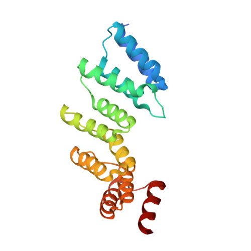
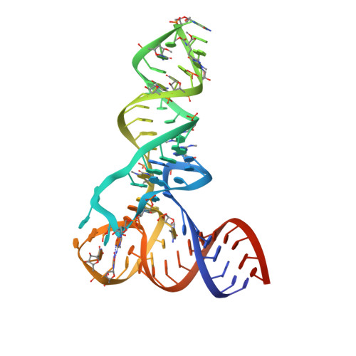
Pentatricopeptide repeats of protein-only RNase P use a distinct mode to recognize conserved bases and structural elements of pre-tRNA.

Teramoto, T.Kaitany, K.J.Kakuta, Y.Kimura, M.Fierke, C.A.Hall, T.M.T.

(2020) Nucleic Acids Res 48: 11815-11826

  • DOI: https://doi.org/10.1093/nar/gkaa627
  • Primary Citation of Related Structures:  
    6LVR

  • PubMed Abstract: 

    Pentatricopeptide repeat (PPR) motifs are α-helical structures known for their modular recognition of single-stranded RNA sequences with each motif in a tandem array binding to a single nucleotide. Protein-only RNase P 1 (PRORP1) in Arabidopsis thaliana is an endoribonuclease that uses its PPR domain to recognize precursor tRNAs (pre-tRNAs) as it catalyzes removal of the 5'-leader sequence from pre-tRNAs with its NYN metallonuclease domain. To gain insight into the mechanism by which PRORP1 recognizes tRNA, we determined a crystal structure of the PPR domain in complex with yeast tRNAPhe at 2.85 Å resolution. The PPR domain of PRORP1 bound to the structurally conserved elbow of tRNA and recognized conserved structural features of tRNAs using mechanisms that are different from the established single-stranded RNA recognition mode of PPR motifs. The PRORP1 PPR domain-tRNAPhe structure revealed a conformational change of the PPR domain upon tRNA binding and moreover demonstrated the need for pronounced overall flexibility in the PRORP1 enzyme conformation for substrate recognition and catalysis. The PRORP1 PPR motifs have evolved strategies for protein-tRNA interaction analogous to tRNA recognition by the RNA component of ribonucleoprotein RNase P and other catalytic RNAs, indicating convergence on a common solution for tRNA substrate recognition.


  • Organizational Affiliation

    Epigenetics and Stem Cell Biology Laboratory, National Institute of Environmental Health Sciences, National Institutes of Health, Research Triangle Park, NC 27709, USA.


Macromolecules

Find similar proteins by:  (by identity cutoff)  |  3D Structure
Entity ID: 1
MoleculeChains Sequence LengthOrganismDetailsImage
Proteinaceous RNase P 1, chloroplastic/mitochondrialA [auth C],
B [auth A]
208Arabidopsis thalianaMutation(s): 3 
Gene Names: PRORP1At2g32230F22D22.2
EC: 3.1.26.5
UniProt
Find proteins for Q66GI4 (Arabidopsis thaliana)
Explore Q66GI4 
Go to UniProtKB:  Q66GI4
Entity Groups  
Sequence Clusters30% Identity50% Identity70% Identity90% Identity95% Identity100% Identity
UniProt GroupQ66GI4
Sequence Annotations
Expand
  • Reference Sequence
Find similar nucleic acids by:  (by identity cutoff)  |  3D Structure
Entity ID: 2
MoleculeChains LengthOrganismImage
yeast phenylalanine tRNAC [auth D],
D [auth B]
76Saccharomyces cerevisiae
Sequence Annotations
Expand
  • Reference Sequence
Experimental Data & Validation

Experimental Data

  • Method: X-RAY DIFFRACTION
  • Resolution: 2.85 Å
  • R-Value Free: 0.255 
  • R-Value Work: 0.235 
  • Space Group: P 21 21 2
Unit Cell:
Length ( Å )Angle ( ˚ )
a = 84.48α = 90
b = 131.701β = 90
c = 155.556γ = 90
Software Package:
Software NamePurpose
REFMACrefinement
HKL-2000data reduction
HKL-2000data scaling
PHASERphasing

Structure Validation

View Full Validation Report


Entry History & Funding Information

Deposition Data


Funding OrganizationLocationGrant Number
National Institutes of Health/National Institute of Environmental Health Sciences (NIH/NIEHS)United States1ZIA50165

Revision History  (Full details and data files)

  • Version 1.0: 2020-08-12
    Type: Initial release
  • Version 1.1: 2020-12-23
    Changes: Database references
  • Version 1.2: 2023-11-29
    Changes: Data collection, Database references, Derived calculations, Refinement description