


The crystal structure of diamondback moth ryanodine receptor SPRY1 domain
Nayak, B., Zhou, Y., Salauddin, N., Lin, L., You, M., You, S., Yuchi, Z.To be published.
Experimental Data Snapshot
Starting Model: experimental
View more details
Entity ID: 1 | |||||
|---|---|---|---|---|---|
| Molecule | Chains | Sequence Length | Organism | Details | Image |
| Ryanodine receptor 1 | A [auth F], B | 195 | Plutella xylostella | Mutation(s): 2 | 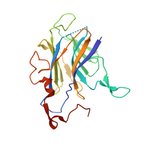 |
UniProt | |||||
Entity Groups | |||||
| Sequence Clusters | 30% Identity50% Identity70% Identity90% Identity95% Identity100% Identity | ||||
| UniProt Group | G8EME3 | ||||
Sequence AnnotationsExpand | |||||
Reference Sequence | |||||
| Ligands 2 Unique | |||||
|---|---|---|---|---|---|
| ID | Chains | Name / Formula / InChI Key | 2D Diagram | 3D Interactions | |
| GOL (Subject of Investigation/LOI) Download:Ideal Coordinates CCD File | C [auth F], E [auth B], F [auth B], G [auth B] | GLYCEROL C3 H8 O3 PEDCQBHIVMGVHV-UHFFFAOYSA-N |  | ||
| NA (Subject of Investigation/LOI) Download:Ideal Coordinates CCD File | D [auth F], H [auth B] | SODIUM ION Na FKNQFGJONOIPTF-UHFFFAOYSA-N |  | ||
| Length ( Å ) | Angle ( ˚ ) |
|---|---|
| a = 64.893 | α = 90 |
| b = 64.893 | β = 90 |
| c = 95.767 | γ = 90 |
| Software Name | Purpose |
|---|---|
| PHENIX | refinement |
| HKL-2000 | data scaling |
| PDB_EXTRACT | data extraction |
| HKL-3000 | data reduction |
| PHENIX | phasing |
| Funding Organization | Location | Grant Number |
|---|---|---|
| Ministry of Science and Technology (MoST, China) | China | 2017YFD0201400 |
| Ministry of Science and Technology (MoST, China) | China | 2017YFD0201403 |


