6LM1 | pdb_00006lm1

The crystal structure of cyanorhodopsin (CyR) N4075R from cyanobacteria Tolypothrix sp. NIES-4075


Experimental Data Snapshot

  • Method: X-RAY DIFFRACTION
  • Resolution: 1.90 Å
  • R-Value Free: 
    0.204 (Depositor), 0.208 (DCC) 
  • R-Value Work: 
    0.165 (Depositor), 0.168 (DCC) 
  • R-Value Observed: 
    0.166 (Depositor) 

Starting Model: experimental
View more details

wwPDB Validation 3D Report Full Report

Validation slider image for 6LM1

Ligand Structure Quality Assessment 


This is version 1.2 of the entry. See complete history

Literature

A unique clade of light-driven proton-pumping rhodopsins evolved in the cyanobacterial lineage.

Hasegawa, M.Hosaka, T.Kojima, K.Nishimura, Y.Nakajima, Y.Kimura-Someya, T.Shirouzu, M.Sudo, Y.Yoshizawa, S.

(2020) Sci Rep 10: 16752-16752

  • DOI: https://doi.org/10.1038/s41598-020-73606-y
  • Primary Citation Related Structures: 
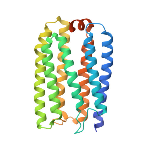
    6LM0, 6LM1

  • PubMed Abstract: 

    Microbial rhodopsin is a photoreceptor protein found in various bacteria and archaea, and it is considered to be a light-utilization device unique to heterotrophs. Recent studies have shown that several cyanobacterial genomes also include genes that encode rhodopsins, indicating that these auxiliary light-utilizing proteins may have evolved within photoautotroph lineages. To explore this possibility, we performed a large-scale genomic survey to clarify the distribution of rhodopsin and its phylogeny. Our surveys revealed a novel rhodopsin clade, cyanorhodopsin (CyR), that is unique to cyanobacteria. Genomic analysis revealed that rhodopsin genes show a habitat-biased distribution in cyanobacterial taxa, and that the CyR clade is composed exclusively of non-marine cyanobacterial strains. Functional analysis using a heterologous expression system revealed that CyRs function as light-driven outward H + pumps. Examination of the photochemical properties and crystal structure (2.65 Å resolution) of a representative CyR protein, N2098R from Calothrix sp. NIES-2098, revealed that the structure of the protein is very similar to that of other rhodopsins such as bacteriorhodopsin, but that its retinal configuration and spectroscopic characteristics (absorption maximum and photocycle) are distinct from those of bacteriorhodopsin. These results suggest that the CyR clade proteins evolved together with chlorophyll-based photosynthesis systems and may have been optimized for the cyanobacterial environment.


  • Organizational Affiliation
    • Atmosphere and Ocean Research Institute, The University of Tokyo, Chiba, 277-8564, Japan.

Macromolecule Content 

  • Total Structure Weight: 93.92 kDa 
  • Atom Count: 6,037 
  • Modeled Residue Count: 678 
  • Deposited Residue Count: 777 
  • Unique protein chains: 1

Macromolecules

Find similar proteins by:|  3D Structure
Entity ID: 1
MoleculeChains  Sequence LengthOrganismDetailsImage
Rhodopsin
A, B, C
259Tolypothrix sp. NIES-4075Mutation(s): 0 
Gene Names: NIES4075_41530
UniProt
Find proteins for A0ACD6B9U0 (Tolypothrix sp. NIES-4075)
Explore A0ACD6B9U0 
Go to UniProtKB:  A0ACD6B9U0
Entity Groups
Sequence Clusters30% Identity50% Identity70% Identity90% Identity95% Identity100% Identity
UniProt GroupA0ACD6B9U0
Sequence Annotations
Expand
Reference Sequence

Small Molecules

Ligands 9 Unique
IDChains Name / Formula / InChI Key2D Diagram3D Interactions
RET
(Subject of Investigation/LOI)

Query on RET


Download:Ideal Coordinates CCD File
CA [auth B],
D [auth A],
WA [auth C]
RETINAL
C20 H28 O
NCYCYZXNIZJOKI-OVSJKPMPSA-N
R16
(Subject of Investigation/LOI)

Query on R16


Download:Ideal Coordinates CCD File
AA [auth A]
OB [auth C]
PB [auth C]
QB [auth C]
UA [auth B]
AA [auth A],
OB [auth C],
PB [auth C],
QB [auth C],
UA [auth B],
W [auth A],
X [auth A],
Y [auth A],
Z [auth A]
HEXADECANE
C16 H34
DCAYPVUWAIABOU-UHFFFAOYSA-N
C14
(Subject of Investigation/LOI)

Query on C14


Download:Ideal Coordinates CCD File
BA [auth A],
TB [auth C],
VA [auth B]
TETRADECANE
C14 H30
BGHCVCJVXZWKCC-UHFFFAOYSA-N
D12
(Subject of Investigation/LOI)

Query on D12


Download:Ideal Coordinates CCD File
NB [auth C]
RA [auth B]
SA [auth B]
TA [auth B]
U [auth A]
NB [auth C],
RA [auth B],
SA [auth B],
TA [auth B],
U [auth A],
V [auth A]
DODECANE
C12 H26
SNRUBQQJIBEYMU-UHFFFAOYSA-N
D10
(Subject of Investigation/LOI)

Query on D10


Download:Ideal Coordinates CCD File
PA [auth B],
QA [auth B],
S [auth A],
T [auth A]
DECANE
C10 H22
DIOQZVSQGTUSAI-UHFFFAOYSA-N
OCT
(Subject of Investigation/LOI)

Query on OCT


Download:Ideal Coordinates CCD File
IB [auth C]
JA [auth B]
JB [auth C]
K [auth A]
KA [auth B]
IB [auth C],
JA [auth B],
JB [auth C],
K [auth A],
KA [auth B],
KB [auth C],
L [auth A],
LA [auth B],
LB [auth C],
M [auth A],
MA [auth B],
MB [auth C],
N [auth A],
NA [auth B],
O [auth A],
OA [auth B],
P [auth A],
Q [auth A],
R [auth A]
N-OCTANE
C8 H18
TVMXDCGIABBOFY-UHFFFAOYSA-N
HEX
(Subject of Investigation/LOI)

Query on HEX


Download:Ideal Coordinates CCD File
AB [auth C]
BB [auth C]
CB [auth C]
DB [auth C]
EA [auth B]
AB [auth C],
BB [auth C],
CB [auth C],
DB [auth C],
EA [auth B],
EB [auth C],
F [auth A],
FA [auth B],
FB [auth C],
G [auth A],
GA [auth B],
GB [auth C],
H [auth A],
HA [auth B],
HB [auth C],
I [auth A],
IA [auth B],
J [auth A],
YA [auth C],
ZA [auth C]
HEXANE
C6 H14
VLKZOEOYAKHREP-UHFFFAOYSA-N
NO3
(Subject of Investigation/LOI)

Query on NO3


Download:Ideal Coordinates CCD File
RB [auth C],
SB [auth C]
NITRATE ION
N O3
NHNBFGGVMKEFGY-UHFFFAOYSA-N
MG
(Subject of Investigation/LOI)

Query on MG


Download:Ideal Coordinates CCD File
DA [auth B],
E [auth A],
XA [auth C]
MAGNESIUM ION
Mg
JLVVSXFLKOJNIY-UHFFFAOYSA-N

Experimental Data & Validation

Experimental Data

  • Method: X-RAY DIFFRACTION
  • Resolution: 1.90 Å
  • R-Value Free:  0.204 (Depositor), 0.208 (DCC) 
  • R-Value Work:  0.165 (Depositor), 0.168 (DCC) 
  • R-Value Observed: 0.166 (Depositor) 
Space Group: C 2 2 21
Unit Cell:
Length ( Å )Angle ( ˚ )
a = 107.92α = 90
b = 129.666β = 90
c = 127.626γ = 90
Software Package:
Software NamePurpose
PHENIXrefinement
XDSdata reduction
Aimlessdata scaling
PHASERphasing

Structure Validation

View Full Validation Report


Ligand Structure Quality Assessment 


Entry History 

Deposition Data

Revision History  (Full details and data files)

  • Version 1.0: 2020-10-21
    Type: Initial release
  • Version 1.1: 2023-11-22
    Changes: Data collection, Database references, Refinement description
  • Version 1.2: 2024-11-06
    Changes: Structure summary