6LII | pdb_00006lii

A quinone oxidoreductase


Experimental Data Snapshot

  • Method: X-RAY DIFFRACTION
  • Resolution: 2.30 Å
  • R-Value Free: 
    0.203 (Depositor), 0.199 (DCC) 
  • R-Value Work: 
    0.180 (Depositor), 0.180 (DCC) 
  • R-Value Observed: 
    0.186 (Depositor) 

Starting Model: experimental
View more details

wwPDB Validation 3D Report Full Report

Validation slider image for 6LII

This is version 1.2 of the entry. See complete history

Literature

Structural insights into vesicle amine transport-1 (VAT-1) as a member of the NADPH-dependent quinone oxidoreductase family.

Kim, S.-Y.Mori, T.Chek, M.F.Furuya, S.Matsumoto, K.Yajima, T.Ogura, T.Hakoshima, T.

(2021) Sci Rep 11: 2120-2120

  • DOI: https://doi.org/10.1038/s41598-021-81409-y
  • Primary Citation Related Structures: 
    6LHR, 6LII

  • PubMed Abstract: 

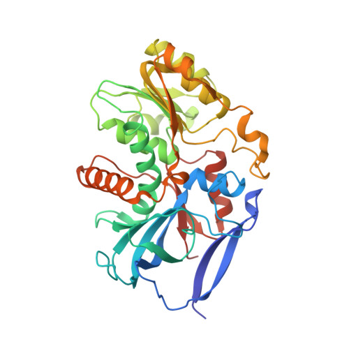
    Vesicle amine transport protein-1 (VAT-1) has been implicated in the regulation of vesicular transport, mitochondrial fusion, phospholipid transport and cell migration, and is a potential target of anticancer drugs. Little is known about the molecular function of VAT-1. The amino acid sequence indicates that VAT-1 belongs to the quinone oxidoreductase subfamily, suggesting that VAT-1 may possess enzymatic activity in unknown redox processes. To clarify the molecular function of VAT-1, we determined the three-dimensional structure of human VAT-1 in the free state at 2.3 Å resolution and found that VAT-1 forms a dimer with the conserved NADPH-binding cleft on each protomer. We also determined the structure of VAT-1 in the NADP-bound state at 2.6 Å resolution and found that NADP binds the binding cleft to create a putative active site with the nicotine ring. Substrate screening suggested that VAT-1 possesses oxidoreductase activity against quinones such as 1,2-naphthoquinone and 9,10-phenanthrenequinone.


  • Organizational Affiliation
    • Structural Biology Laboratory, Nara Institute of Science and Technology, 8916-5 Takayama, Ikoma, Nara, 630-0192, Japan.

Macromolecule Content 

  • Total Structure Weight: 152.22 kDa 
  • Atom Count: 10,545 
  • Modeled Residue Count: 1,372 
  • Deposited Residue Count: 1,412 
  • Unique protein chains: 1

Macromolecules

Find similar proteins by:|  3D Structure
Entity ID: 1
MoleculeChains  Sequence LengthOrganismDetailsImage
Synaptic vesicle membrane protein VAT-1 homolog
A, B, C, D
353Homo sapiensMutation(s): 0 
Gene Names: VAT1
EC: 1.6.5.5
UniProt & NIH Common Fund Data Resources
Find proteins for Q99536 (Homo sapiens)
Explore Q99536 
Go to UniProtKB:  Q99536
PHAROS:  Q99536
GTEx:  ENSG00000108828 
Entity Groups
Sequence Clusters30% Identity50% Identity70% Identity90% Identity95% Identity100% Identity
UniProt GroupQ99536
Sequence Annotations
Expand
Reference Sequence

Experimental Data & Validation

Experimental Data

  • Method: X-RAY DIFFRACTION
  • Resolution: 2.30 Å
  • R-Value Free:  0.203 (Depositor), 0.199 (DCC) 
  • R-Value Work:  0.180 (Depositor), 0.180 (DCC) 
  • R-Value Observed: 0.186 (Depositor) 
Space Group: P 32 2 1
Unit Cell:
Length ( Å )Angle ( ˚ )
a = 69.349α = 90
b = 69.349β = 90
c = 581.807γ = 120
Software Package:
Software NamePurpose
PHENIXrefinement
PHENIXrefinement
XDSdata reduction
XDSdata scaling
PHASERphasing

Structure Validation

View Full Validation Report


Entry History 

Deposition Data

Revision History  (Full details and data files)

  • Version 1.0: 2021-01-20
    Type: Initial release
  • Version 1.1: 2021-02-10
    Changes: Database references
  • Version 1.2: 2023-11-22
    Changes: Data collection, Database references, Refinement description